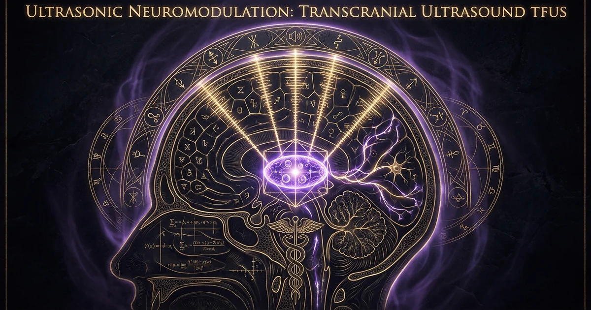
Ultrasonic Neuromodulation: Transcranial Ultrasound TFUS
Executive Summary & Theoretical Thesis: The Acoustic Neuromodulation Paradigm
Spatial Resolution Limits of Electromagnetic vs. Acoustic Modalities
Contemporary clinical and experimental neurotechnologies have long contended with the physical trade-off between spatial invasiveness and anatomical focal depth. Modalities relying on macroscopic electromagnetic fields, specifically Transcranial Magnetic Stimulation (TMS) and transcranial Electrical Stimulation (tES, encompassing tDCS and tACS), are fundamentally constrained by Laplace’s equation governing quasistatic potential distributions within volume conductors. As electromagnetic flux crosses the high-impedance boundary of the calvarium, the primary electric field undergoes pronounced spatial dispersion, acting as a low-pass spatial filter. Consequently, achieving focal depolarization at subcortical targets—such as the centromedian thalamic nuclei, the subthalamic nucleus, or the basolateral amygdala—is physically impossible via conventional surface coils without exerting suprathreshold field intensities upon intervening neocortical circuits.
In contrast, transcranial focused ultrasound neuromodulation tfus deep brain protocols exploit high-frequency acoustic wave propagation, which does not decay under the electrostatic boundary constraints of a dielectric-field. By converting high-frequency electrical signals into coherent longitudinal-waves via piezoelectric ceramic arrays, acoustic beams propagate through human cranial architecture with millimetric wavelengths ($\lambda \approx 1.5$ to $3.0\text{ mm}$ at $0.5\text{ to }1.0\text{ MHz}$). Because acoustic impedance mismatches between soft tissue layers are orders of magnitude lower than their electrical impedance differentials, acoustic waves retain spatial coherence, allowing convergent beam trajectories to form a focused acoustic ellipsoid at arbitrary depths within the brain volume.
Transcranial Magnetic/Electrical Stimulation (TMS/tDCS)
- Penetration Depth: Limited to superficial neocortex ($\approx 15\text{–}25\text{ mm}$); subcortical reach requires indiscriminate cortical stimulation.
- Lateral Spatial Resolution: Diffuse, typically spanning $10\text{–}30\text{ mm}$; constrained by the spatial low-pass filtering of the cranium.
- Skull Attenuation Dynamics: High trans-cranial electrical impedance attenuates current density, causing lateral field shunting through the scalp.
- Biophysical Mechanism: Macroscopic electric field gradient alters neuronal resting membrane potential via capacitive charge transfer along the axodendritic axis.
Transcranial Focused Ultrasound (tFUS)
- Penetration Depth: Transcranial reach spans entire human neuroanatomy ($>120\text{ mm}$), accessing deep nuclei with equal efficacy.
- Lateral Spatial Resolution: Millimetric focal envelope ($1.0\text{–}3.0\text{ mm}$ lateral, $5.0\text{–}10.0\text{ mm}$ axial), operating beyond optical or magnetic focal limits.
- Skull Attenuation Dynamics: High acoustic absorption and phase aberration; mitigated via sub-megahertz frequencies and multi-element CT phase conjugation.
- Biophysical Mechanism: Acoustic radiation force, nanoscale intramembrane cavitation, and mechanosensitive ion channel gating under non-thermal pulsing.
Mechanotransductive Gating of Deep-Brain Connectomics
The clinical reality of non-invasive deep brain stimulation relies upon translating mechanical pressure gradients directly into cellular neurochemical signaling. When acoustic energy concentrates within a target voxel, the local tissue microenvironment is subjected to cyclic variations in compressive and tensile stress. This dynamic biomechanical state alters the physical conformation of the cellular membrane, establishing an unconventional pathway for modulating deep-brain connectomics without the electrochemical invasiveness of surgically implanted deep brain stimulation (DBS) electrodes.
The mechanism by which tFUS engages these deep neural networks is fundamentally mechanotransductive. Rather than acting as an external dipole that forces passive current flow through a resistive extracellular matrix, tFUS perturbs the lipid bilayer’s tension state. This mechanical alteration initiates a cascade of intracellular signaling events, modulates local field potentials, and resets aberrant oscillopathies within deep cortico-striatal-thalamic loops. Through these mechanodynamic vectors, non-invasive deep brain stimulation transitions from a theoretical ideal into a reproducible biophysical modality capable of selectively modulating basal ganglia output, interrupting epileptic foci, or altering limbic affective valuation nodes.
The Acoustic Radiation Force Vector in Non-Thermal Regimes
A foundational distinction in therapeutic ultrasound is the operational divergence between high-intensity focused ultrasound (HIFU), optimized for targeted thermal ablation, and low-intensity low-frequency ultrasound (LILFU), optimized for non-destructive neuroregulation. In neuromodulatory regimes, acoustic parameters are tuned to suppress bulk tissue temperature elevation ($\Delta T < 0.1^\circ\text{C}$), operating far below the threshold for thermal protein denaturation or microvascular coagulation.
Under these non-thermal regimes, bioeffects are governed by the acoustic radiation force (ARF) vector. Acoustic radiation force originates from the transfer of linear momentum from the propagating acoustic wave to the absorbing and scattering viscoelastic medium of the brain parenchyma. This momentum transfer induces a directional mechanical body force within the focal envelope:
$$\mathbf{F}_v = \frac{2\alpha \mathbf{I}}{c}$$
where $\alpha$ is the acoustic absorption coefficient of the neural tissue, $\mathbf{I}$ is the cycle-averaged acoustic intensity vector, and $c$ is the longitudinal speed of sound in the parenchymal medium. This steady mechanical displacement field imparts strain along neuronal membranes and glial processes, displacing cytoskeletal elements and triggering mechanosensitive gating cascades without relying on the scalar thermodynamic shifts characteristic of ablative neurosurgery.
Historical Lineage & Experimental Precedents: From High-Intensity Lesions to Non-Thermal Pulsing
The Fry Brothers’ Laboratory and Early Neurosonology (1950s)
The biophysical intersection of high-frequency acoustics and the central nervous system was first rigorously mapped in the late 1940s and 1950s at the Bioacoustics Research Laboratory of the University of Illinois by William J. Fry and Francis J. Fry. The Fry brothers recognized that convergent multi-beam ultrasound could focus non-ionizing mechanical energy at designated subcortical coordinates with millimetric precision. However, these pioneering protocols were technologically restricted by early mid-century transducer materials and the complete absence of non-invasive calvarial phase-aberration correction.
Consequently, early neurosonology required craniectomies—surgically removing acoustic windows in animal models and human parkinsonian patients—to eliminate the massive acoustic attenuation, scattering, and focal degradation caused by the calvarium. These historical trials concentrated almost exclusively on high acoustic energy regimes designed to generate targeted, irreversible structural lesions within the ansa lenticularis and substantia nigra to alleviate tremors.
Despite this ablative imperative, William Fry recorded anomalous physiological observations: when acoustic energy levels were titrated to sub-ablative thresholds, neural conduction through visual and motor pathways could be transiently suspended and subsequently recovered without detectable histopathological destruction.
“It is possible to produce localized reversible changes in the central nervous system by the proper choice of the physical parameters of the ultrasound: frequency, sound levels, and duration of exposure… Functional recovery can occur with no gross or histological signs of irreversible damage.” — Fry, W. J., et al. (1958). Science, 127(3289), 83-84.
This observation serves as the foundational empirical antecedent to low-intensity ultrasound neuromodulation, anticipating the clinical transition from mechanical tissue ablation to reversible mechanosensitive signaling—a milestone later brought to trans-calvarial clinical utility via CT-based acoustic phase correction (Hynynen, US Patent 6,428,477).
The Paradigm Shift to Low-Intensity Low-Frequency Ultrasound (LILFU)
Decades after Fry’s observations, the realization that acoustic radiation could induce functional, reversible electrophysiological transitions led to a modern resurgence in ultrasonic neuromodulation. The transition toward low-intensity, low-frequency ultrasound (LILFU)—typically defined as spatial-peak temporal-average intensities ($I_{spta}$) below $720\text{ mW/cm}^2$ and frequencies between $0.25\text{ and }0.7\text{ MHz}$—demonstrated that acoustic pulses modulate intrinsic neuronal firing patterns.
Seminal work by Tufail et al. (2010) demonstrated that pulsed acoustic wave-trains delivered through intact rodent crania evoked motor responses, induced local field potential (LFP) shifts, and synchronized cortical and subcortical oscillatory networks without elevating parenchymal temperature. Concurrently, investigations led by W. Jamie Tyler contextualized these results within contemporary molecular mechanics, demonstrating that non-cavitational low-pressure waves drive the opening of both voltage-gated and stretch-activated ion channels. This shifted neurosonology away from thermal destruction and toward reversible biophysical computation.
Acoustic Modality Parameter Divergence:
┌────────────────────────┬─────────────────────────┬─────────────────────────┐
│ Metric / Domain        │ Ablative HIFU           │ Neuromodulatory tFUS    │
├────────────────────────┼─────────────────────────┼─────────────────────────┤
│ Frequency Range        │ 0.65 – 1.5 MHz          │ 0.25 – 0.70 MHz         │
│ Intensity (Ispta)      │ > 1000 W/cm²            │ 0.1 – 3.0 W/cm²         │
│ Duty Cycle             │ Continuous (100%)       │ Pulsed (0.5% – 10%)     │
│ Parenchymal Temp Shift │ ΔT > 20°C (Coagulation) │ ΔT < 0.1°C (Isothermal) │
│ Primary Bio-Effect     │ Thermal Denaturation    │ Mechanotransduction     │
└────────────────────────┴─────────────────────────┴─────────────────────────┘
CT-Derived Phased-Array Transducers and Transcranial De-Aberration
The clinical translation of non-invasive trans-calvarial acoustic delivery required overcoming the irregular geometry and variable bone mineral density of the human calvarium. The human skull acts as an inhomogeneous acoustic plate; variations in thickness and cortical-trabecular ratios across the frontal, parietal, and temporal bones generate severe phase distortions, wavefront scattering, and acoustic refraction. When an uncorrected spherical acoustic wavefront passes through these heterogeneities, the focal point defocuses, shifts in three-dimensional space, and loses the acoustic intensity density required to exceed biological response thresholds.
This engineering barrier was overcome through the development of multi-element hemispherical phased-array transducers integrated with high-resolution computed tomography (CT) registration algorithms. By mapping the Hounsfield units (HU) of a patient’s skull to microstructural bone density and acoustic velocity distributions, finite-difference time-domain (FDTD) simulations compute the precise acoustic phase shift introduced by every square millimeter of calvarial bone. Each element within a multi-element array (typically comprising 1024 or more independent piezoceramic channels) is programmed with an inverse phase offset. The emitted wavelets thus undergo constructive interference precisely at the intended stereotactic focal coordinate within the subcortical target, rendering the cranium acoustically transparent via digital phase de-aberration.
Mathematical Formalism & Physical Mechanics: Acoustic Radiation Force, Cavitation, and Membrane Electrophysiology
Kuznetsov-Westervelt Non-Linear Acoustic Wave Equation
The propagation of high-amplitude ultrasound waves through lossy, inhomogeneous, viscoelastic biological media departs from classical linear acoustics. To accurately compute the focal volume, diffraction patterns, and higher-order harmonic generation within brain tissue, acoustic propagation is modeled via the non-linear Kuznetsov-Westervelt equation:
$$\nabla^2 p - \frac{1}{c_0^2}\frac{\partial^2 p}{\partial t^2} + \frac{\delta}{c_0^4}\frac{\partial^3 p}{\partial t^3} + \frac{\beta}{\rho_0 c_0^4}\frac{\partial^2 p^2}{\partial t^2} = 0$$
In this formulation:
- $p$ represents the acoustic sound pressure distribution,
- $c_0$ denotes the small-signal longitudinal speed of sound in the parenchyma ($\approx 1540\text{ m/s}$),
- $\rho_0$ is the ambient equilibrium mass density of the tissue ($\approx 1040\text{ kg/m}^3$),
- $\delta$ denotes the acoustic diffusivity of the medium, parameterizing thermoviscous dissipation: $$\delta = \frac{1}{\rho_0}\left[\frac{4}{3}\mu + \mu_B + \kappa\left(\frac{1}{c_v} - \frac{1}{c_p}\right)\right]$$ where $\mu$ is shear viscosity, $\mu_B$ is bulk viscosity, and $\kappa$ is thermal conductivity,
- $\beta = 1 + \frac{B}{2A}$ is the parameter of non-linearity, where $B/A$ is the ratio of non-linear to linear pressure coefficients of the biological substrate.
At typical neuromodulatory intensities, non-linear harmonic distortion transfers energy from the fundamental operational frequency $f_0$ into higher harmonic modes ($2f_0, 3f_0, \dots$). These higher modes undergo accelerated attenuation within the deep focal envelope, steepening the localized acoustic radiation force gradient and narrowing the effective lateral half-maximum intensity dimensions of the focal spot.
Bilayer Sonophore Model and Intramembrane Cavitation Mechanics
The primary biophysical mechanism translating non-thermal acoustic pressure oscillations into cellular electrical excitation is described by the Bilayer Sonophore (BLS) model formulated by Krasovitski, Frenkel, Shoham, and Kimmel (2011). Biological membranes consist of an antiparallel leaflet array of amphiphilic phospholipids separated by an intramembrane hydrophobic core. The BLS model posits that under the tensile phase of a low-frequency ultrasound wave, negative acoustic pressure ($P_{\text{neg}}$) induces intramembrane-cavitation: the reversible mechanical separation of the two lipid leaflets on a sub-nanometer scale.
Bilayer Sonophore (BLS) Intramembrane Cavitation Dynamic:

 Resting State (Zero Acoustic Pressure)
   Extracellular Fluid
   ===========================  Top Phospholipid Leaflet
   ---------------------------  Hydrophobic Core (d_0 ≈ 3.5 nm)
   ===========================  Bottom Phospholipid Leaflet
   Intracellular Cytoplasm

 Peak Rarefaction (Negative Acoustic Pressure: P_neg)
   Extracellular Fluid
      /~~~~~~~~~~~~~~~~~\       Outward Leaflet Deflection
     |   Nanoscale Void  |      Intramembrane Cavity (Z-expansion)
      \~~~~~~~~~~~~~~~~~/       Inward Leaflet Deflection
   Intracellular Cytoplasm
   [ΔC_m Shift -> Discharges Membrane Capacitance -> Depolarization]
As the local pressure oscillates between positive compression and negative rarefaction, the intramembrane space acts as an acoustic energy harvester. The dynamic displacement of the leaflet separation, $Z(t)$, is governed by balancing the localized internal gas pressure, the static hydrophobic surface tension, the surrounding viscoelastic resistance of the cytomatrix, and the driving acoustic wave:
$$m_s \ddot{Z} + b_s \dot{Z} + \frac{2\gamma}{Z} + \Pi_{\text{visco}}(Z, \dot{Z}) = -P_A(t)$$
where $m_s$ is the effective leaflet areal mass density, $b_s$ represents the viscous damping coefficient of the surrounding lipid-water interface, $\gamma$ is the interfacial surface tension, and $P_A(t)$ is the incoming non-linear acoustic pressure profile.
Because the electrical capacitance of a biological membrane per unit area is inversely proportional to the dielectric thickness $d$ separating the extracellular and intracellular electrolyte pools:
$$C_m(t) = \frac{\epsilon_0 \epsilon_r}{d(t)}$$
the rapid, cyclic expansion and contraction of the intramembrane core induces large, non-linear shifts in membrane capacitance ($\frac{dC_m}{dt}$). Under the biological conservation of transmembrane charge ($Q = C_m V_m$), this mechanical capacitance modulation generates a capacitive displacement current:
$$I_C = C_m \frac{dV_m}{dt} + V_m \frac{dC_m}{dt}$$
This displacement current shifts the resting membrane potential ($V_m$) toward depolarizing levels. Once the threshold is crossed, it activates voltage-gated sodium channels and fires genuine all-or-nothing axonal action potentials.
The primary body force density $\mathbf{F}_v$ transferred to the targeted parenchymal matrix is computed via the spatial attenuation of the time-averaged acoustic Poynting vector:
$$\mathbf{F}v = \frac{2 \alpha \langle \mathbf{I} \rangle}{c} = \frac{\alpha p{\text{peak}}^2}{\rho_0 c^2}$$
To ensure this radiation force remains within the non-destructive, non-cavitational neuroregulatory window, experimental and clinical exposures must be quantified relative to the dimensionless Mechanical Index (MI):
$$\text{MI} = \frac{P_{\text{neg}}}{\sqrt{f_0}}$$
where $P_{\text{neg}}$ is the peak rarefactional pressure derated for in situ tissue loss (expressed in MPa), and $f_0$ is the acoustic fundamental frequency (expressed in MHz). For human neuromodulation regimes:
- The target window for pure mechanotransductive non-thermal gating is constrained to: $0.2 \le \text{MI} \le 0.7$.
- The critical threshold for inertial cavitation in the absence of exogenous contrast microbubbles occurs at: $\text{MI}_{\text{crit}} \approx 1.9$. Exceeding this threshold causes rapid bubble collapse, shock-wave generation, and mechanical parenchymal shearing.
Piezoelectric and Mechanosensitive Ion Channel Gating Dynamics
Beyond intramembrane capacitance shifts, modulating synaptic plasticity sound protocols mechanically engage membrane-bound mechanosensitive ion channels. The mechanical strain imparted by both the dynamic acoustic radiation force vector and the localized leaflet strain directly alters lateral membrane tension ($\sigma_{\text{mem}}$), calculated via Laplace’s law applied to the local bilayer curvature:
$$\Delta \sigma_{\text{mem}} = \frac{\Delta P_{\text{trans}} \cdot R_{\text{curv}}}{2}$$
This acoustic tension activates several primary classes of mechanosensitive ion channel proteins:
- PIEZO1 and PIEZO2 Complexes: These large, trimeric mechanosensitive channels possess an expansive, curved transmembrane propeller domain. When acoustic radiation force increases lateral membrane tension, the propeller-like blade flattens within the lipid plane, opening the central conduction pore to drive an inward flux of $\text{Ca}^{2+}$ and $\text{Na}^+$ ions.
- Two-Pore Domain Potassium Channels (K2P: TREK-1, TRAAK): These hyperpolarizing channels respond to negative membrane curvature and lateral stretch. Mechanical membrane tension modulates the conformational equilibrium of the channel’s transmembrane helices, regulating background outward potassium currents and providing a direct mechanism for acoustic neuronal suppression.
- Transient Receptor Potential Channels (TRPV4, TRPC1): These channels are tethered to the cortical cytoskeleton and respond to acoustic shear strain. Their activation induces long-duration intracellular calcium oscillations that trigger downstream transcriptional cascades.
This multi-channel mechanotransduction paradigm shows that the central nervous system’s response to acoustic stimulation is not driven by thermal gradients or electrical capacitive fields, but by the physical activation of mechanosensitive channel kinetics.
Empirical Evidence & Observational Data: Blood-Brain Barrier Transience and Synaptic Plasticity
Acoustic Microbubble Cavitation and Targeted BBB Permeabilization
A primary experimental and clinical application of focused acoustic fields is opening blood brain barrier microbubbles protocols. The non-permeabilized neurovascular unit is maintained by endothelial continuous tight junctions—composed principally of zonula occludens (ZO-1, ZO-2), claudin-5, and occludin—which prevent the paracellular diffusion of macromolecules, therapeutic proteins, and gene vectors larger than approximately 400 Daltons from the systemic circulation into the parenchyma.
Acoustic Microbubble Dynamic within Brain Microvasculature:
Brain Capillary Lumen
════════════════════════════════════════════════════════════════
Endothelial Cell Wall (ZO-1, Claudin-5 Tight Junctions)
-----------------[ Tight Junction Intact ]---------------------
( ( ( Acoustic Wave: f = 0.5 MHz, P_neg = 0.3 MPa ) ) )
╭────────╮
│ Micro- │  <– Stable Non-Inertial Cavitation
│ bubble │      (Symmetric Expansion & Contraction)
╰────────╯
│
▼ Mechanical Push-Pull Shear Stress
-----------------[ Transient Pore Opening ]---------------------
Extravasation of Macromolecules / Neurotherapeutics
════════════════════════════════════════════════════════════════
Brain Parenchyma
When lipid-, polymer-, or protein-shelled perfluorocarbon gas microbubbles (mean diameter: $1.0\text{–}3.0\ \mu\text{m}$) are administered systemically, their acoustic cross-section exceeds their geometric physical cross-section by several orders of magnitude.
Exposing these circulating microbubbles to sub-megahertz transcranial ultrasound focused on target deep nuclei induces stable non-inertial cavitation. The microbubbles undergo sustained, symmetric volumetric oscillations without collapsing. This periodic expansion and contraction applies localized push-pull forces and hydrodynamic micro-streaming against the luminal surface of the brain microvasculature.
These mechanical forces stretch endothelial membranes, downregulate tight-junction scaffold complexes, and stimulate active caveolae-mediated transcellular transport. This targeted permeabilization opens the blood-brain barrier for a transient, safe window of 4 to 24 hours without inducing petechial hemorrhages or persistent neuroinflammatory microgliosis.
“Focal, reversible disruption of the blood-brain barrier can be produced noninvasively using low-intensity pulsed focused ultrasound in combination with a microbubble contrast agent, guided and monitored by magnetic resonance imaging. No evidence of widespread parenchymal damage or tissue necrosis is observed within the target region when applied at calibrated acoustic pressures.” — Hynynen, K., et al. (2001). Radiology, 220(3), 640-646.
Modern replications using multi-electrode arrays confirm that calibrated acoustic cavitation at peak negative pressures between 0.3 and 0.45 MPa enables molecular delivery while keeping the electrophysiological health and functional synaptic transmission of local neural networks intact.
Modulation of Long-Term Potentiation (LTP) and Synaptic Architecture
Beyond the transient depolarization of axonal shafts, modulating synaptic plasticity sound paradigms selectively tune long-term synaptic weightings. The direction of this plasticity—bi-directionally steering neural circuits toward Long-Term Potentiation (LTP) or Long-Term Depression (LTD)—depends on the acoustic pulsing kinetics:
- Pulse Repetition Frequency (PRF): The frequency at which individual ultrasound tone bursts are delivered.
- Duty Cycle (DC): The ratio of pulse duration to total pulse period, which dictates the rate of acoustic radiation force delivery over time.
- Sonication Duration: The overall temporal profile of the acoustic exposure.
Empirical slice electrophysiology and in vivo rodent field recordings reveal that high-frequency pulsing regimes (e.g., $\text{PRF} = 1\text{ kHz}$, $\text{Duty Cycle} = 5\text{–}10%$, sonication duration $\ge 30\text{ seconds}$) induce sustained synaptic potentiation that persists for hours post-sonication. This state mirrors classical tetanus-induced LTP, driven by persistent calcium influx through mechanosensitive PIEZO channels and the unblocking of NMDA receptors via sustained acoustic depolarization. This calcium influx triggers downstream phosphorylation of $\text{Ca}^{2+}$/calmodulin-dependent protein kinase II (CaMKII) and recruits AMPA receptor subunits (GluA1) to the post-synaptic density.
Conversely, continuous low-duty-cycle regimes (e.g., $\text{PRF} \le 10\text{ Hz}$, $\text{Duty Cycle} < 1%$) selectively drive long-term depression (LTD) by inducing low-amplitude calcineurin-dependent protein phosphatase cascades. These downstream cascades alter dendritic spine morphology, reorganize actin microfilaments within post-synaptic boutons, and upregulate the secretion of Brain-Derived Neurotrophic Factor (BDNF) via the TrkB receptor signaling axis. These results establish that acoustic energy serves as a flexible mechanobiological driver of neuroplasticity.
Electrophysiological (fMRI/LFP) Verification in Deep Subcortical Circuits
Verifying that tFUS drives targeted deep-brain responses—rather than diffuse cortical arousal or auditory co-activation artifacts—requires simultaneous multi-modal electrophysiological and functional imaging readouts. Concurrent tFUS-fMRI experiments in non-human primates and human subjects demonstrate spatially constrained blood-oxygen-level-dependent (BOLD) signal modulations localized to deep subcortical structures.
Targeting the ventral posterolateral nucleus (VPL) of the thalamus yields focal BOLD signal transitions restricted to the target thalamic coordinates, alongside functional connectivity modulations across the primary somatosensory cortex (S1). Local field potential (LFP) spectral analysis from stereotactic deep-brain depth electrodes demonstrates that tFUS suppresses aberrant high-frequency oscillations (25–35 Hz beta-band synchronization) in the subthalamic nucleus of parkinsonian models, replicating the therapeutic electrophysiological signature of high-frequency deep brain stimulation (DBS).
Rigorous spatial controls confirm that when the acoustic focus is shifted by as little as $2.0\text{ mm}$ off-target, localized subcortical modulation ceases, validating the spatial fidelity of trans-calvarial focused sound fields.
System Architecture: Phased-Array Transducer Beamforming & Phase Correction
Heterogeneous Calvarial Acoustic Impedance Mapping via High-Res CT
The human skull consists of three distinct layers: an outer cortical bone table, a central trabecular diploë characterized by dynamic porous marrow-filled cancellous architecture, and an inner cortical bone table. The speed of sound through dense cortical bone reaches $c_{\text{bone}} \approx 2800\text{–}3100\text{ m/s}$, compared to the velocity within the brain parenchyma ($c_{\text{brain}} \approx 1540\text{ m/s}$), yielding a dramatic acoustic impedance mismatch:
$$Z_{\text{bone}} = \rho_{\text{bone}} c_{\text{bone}} \approx 6.0 \times 10^6\text{ kg}/(\text{m}^2\cdot\text{s}) \quad \gg \quad Z_{\text{brain}} \approx 1.6 \times 10^6\text{ kg}/(\text{m}^2\cdot\text{s})$$
This impedance mismatch causes substantial reflection at the scalp-skull and skull-dura interfaces, while acoustic speed variations within the heterogeneous diploë layer introduce spatially localized phase delays across the propagating wavefront.
To overcome this calvarial distortion, modern transcranial systems employ high-resolution stereotactic CT scans. The tissue absorption indices (Hounsfield Units, HU) from these scans are segmented and converted into local mass density ($\rho$) and acoustic velocity ($c$) distributions via established empirical calibration profiles:
$$c(\mathbf{r}) = c_{\text{water}} + (c_{\text{max}} - c_{\text{water}}) \left( \frac{\text{HU}(\mathbf{r}) - \text{HU}{\text{water}}}{\text{HU}{\text{max}} - \text{HU}_{\text{water}}} \right)$$
This voxel-level spatial model maps the unique calvarial phase aberration profile of each patient, providing the computational foundation for trans-cranial de-aberration.
Time-Reversal and Phase Conjugation Acoustic Computation
Restoring a diffraction-limited focal envelope within subcortical target structures relies on the computational principles of numerical phase conjugation and time-reversal acoustics. The wave equation in non-dissipative or weakly dissipative media exhibits time-reversal invariance: if a spatiotemporal acoustic field $p(\mathbf{r}, t)$ solves the acoustic wave equation, the time-reversed version $p(\mathbf{r}, -t)$ is also an exact solution.
In clinical practice, this symmetry is exploited through virtual-source acoustic simulations. A virtual point source is computationally positioned at the intended stereotactic target coordinate within the subthalamic nucleus or amygdala. The divergent spherical pressure wave emitted from this virtual source is propagated backward through the heterogeneous skull model using three-dimensional finite-difference time-domain (FDTD) or k-space pseudo-spectral numerical algorithms:
$$\frac{\partial^2 p}{\partial t^2} = -c_0^2 L p$$
where $L$ represents the spatial differential operator accounting for localized calvarial density and elasticity variations.
As the acoustic wave emerges through the calvarium, each individual element of the external hemispherical phased-array transducer captures the phase $\phi_n$ and amplitude $A_n$ of the simulated wave arrival. In actual therapy delivery, the physical transducer array inverts these recorded parameters: each element emits an acoustic wavelet with a phase delay of $-\phi_n$ and an amplitude proportional to $A_n$. As these phase-conjugated waves traverse the skull, the calvarial phase delays are canceled out. The aberrated wave fronts coalesce via constructive interference at the deep intracranial target, forming a precise, un-defocused focal spot.
Thermal Dose Unit (CEM43) Safety Limits and Focal Envelope Localization
To guarantee absolute safety during non-invasive deep brain stimulation, thermal accumulation within the targeted neural tissue and adjacent bone plates must be continuously monitored and mathematically constrained. Thermal accumulation is parameterized using the Cumulative Equivalent Minutes at 43°C (CEM43) metric, formulated by Sapareto and Dewey:
$$\text{CEM43} = \sum_{t=0}^{t_{\text{final}}} R^{(43 - T(t))} \cdot \Delta t$$
where $T(t)$ is the cycle-averaged temperature during the time step $\Delta t$, and $R$ is an empirical parameter ($R = 0.25$ for $T < 43^\circ\text{C}$ and $R = 0.5$ for $T \ge 43^\circ\text{C}$).
In low-intensity neuromodulatory regimes, the acoustic exposure parameters are actively managed so that $\text{CEM43}$ remains identically zero throughout the procedure ($\Delta T < 0.1^\circ\text{C}$ within brain tissue, $\Delta T < 0.5^\circ\text{C}$ at the calvarial inner table). Real-time tracking is maintained via proton resonance frequency shift (PRFS) magnetic resonance thermometry, allowing clinicians to verify focal localization within a 2–3 mm³ volume while ensuring surrounding tissue remains unaffected.
Metaphysical Implications & Unified Synthesis: Cymatic Neurodynamics and Universal Resonances
The Brain as a Viscoelastic Cymatic Resonator
The empirical mechanics of transcranial focused ultrasound reveal a deeper neurobiological truth: the brain is not merely an electrochemical circuit governed by classical cable equations. Rather, neural tissue functions as a viscoelastic phononic medium capable of sustaining and processing complex mechanical wave modes. For over a century, neurobiology has focused almost exclusively on electrical dipoles, treating mechanical pressure transients as secondary epiphenomena. Yet, the high mechanosensitivity of neural structures demonstrates that mechanical state variables are fundamental to neural computation.
Viewed through non-linear acoustics and wave-matter interactions, the mammalian brain operates as an intricate cymatic resonator. Biological tissue matrices—with their complex, heterogeneous distributions of lipid bilayers, neurofilaments, extracellular glycosaminoglycans, and cerebrospinal fluid compartments—support standing waves and acoustic dispersion geometries that reflect the physical dynamics of cymatic-modal-nodes. The spatial organization of subcortical nuclei and their interconnected neural tracts mirrors these structural wave-mode patterns:
Macroscopic Wave-Matter Coupling Hierarchy:

   Acoustic Transducer Input (Longitudinal Acoustic Wavevector)
                         │
                         ▼
   Calvarial Boundary (Heterogeneous Acoustic Shell Formulation)
                         │
                         ▼
   Ventricular Fluid Cavities (Acoustic Cavity Modes & Eigenfrequencies)
                         │
                         ▼
   Viscoelastic Parenchyma (Phononic Dispersion Dynamics)
                         │
                         ▼
   Membrane Phospholipid Bilayers (Nanoscale Intramembrane Cavitation)
                         │
                         ▼
   Mechanosensitive Channels: PIEZO1 / TREK-1 (Conformational Ion Flux)
                         │
                         ▼
   Global Synaptic Plasticity & Connectomic Entrainment (Emergent Consciousness)
At this scale, the localized mechanical pressure fields generated during tFUS reflect the same fundamental principles governing cymatic self-organization in classical continuum mechanics.
Macro-Acoustic Coupling: Longitudinal Mechanics in Biological Structures
At the cellular scale, the propagation of longitudinal-waves through viscoelastic tissue mirrors acoustic levitation and particulate patterning in classical fluids. Just as piezoelectric transducers establish acoustic pressure nodes that trap and align suspended particles in air or water, intracranial acoustic fields generate pressure distributions that pattern suspended biomolecules, reorganize the internal tubulin-microtubule cytoskeleton, and adjust synaptic vesicle docking rates along the active zone.
In these systems, longitudinal acoustic vectors couple into cellular mechanics without requiring external transverse shear forces. This acoustic-biological coupling demonstrates that the central nervous system has evolved to sense and process mechanical wave patterns alongside electrical potentials. The axon is not simply an insulated electrical cable; it is an acoustic waveguide through which mechanical displacement pulses—such as the soliton-like mechanical waves that accompany action potentials—propagate in lockstep with transmembrane ionic currents.
Synthesis of Acoustic Cavitation, Cellular Coherence, and Field Geometries
Synthesizing acoustic cavitation dynamics, cellular mechanotransduction, and macro-scale field geometries reveals an integrated view of neurobiology: the brain functions as a multi-scale, mechanosonic continuum. At the nanoscale, the lipid bilayer undergoes sub-nanometer intramembrane expansion and contraction. At the mesoscale, these fluctuations synchronize populations of mechanosensitive ion channels, generating coherent local field potentials. At the macroscale, multi-beam acoustic interference patterns shape deep-brain functional connectomics.
This mechanosonic perspective bridges physical acoustics, cellular physiology, and field theories. The brain is revealed as a dynamic medium where electrical, mechanical, and structural properties are deeply interconnected. Transcranial focused ultrasound neuromodulation shows that the brain does not operate solely through isolated synaptic chemical exchanges. Rather, it functions as a coherent, resonant system where sound and mechanical tension serve as fundamental organizing forces.
Frequently Asked Questions: Advanced Technical & Biophysical Clarifications
Calvarial Heating Dynamics During Sustained tFUS Protocols
Calvarial bone exhibits an acoustic absorption coefficient ($\alpha \approx 10\text{–}20\text{ dB/cm at }1\text{ MHz}$) that is roughly an order of magnitude higher than that of parenchymal soft tissue ($\alpha \approx 0.6\text{–}0.9\text{ dB/cm at }1\text{ MHz}$). Because absorption scales non-linearly with frequency, operating at multi-megahertz diagnostic frequencies risks causing rapid calvarial heating and thermal osteonecrosis if applied over sustained duty cycles.
Calvarial Thermal Safety: Duty Cycle vs. Heat Dissipation

 43°C ────────────────────────────────── Upper Thermal Safety Threshold
 Temp
  │        Active Sonication      Inter-Pulse Interval
  │           (Heat Influx)         (Perfusion Washout)
  │          ╭──────────────╮
  │         ╱                ╲                ╭──────────────╮
  │        ╱                  ╲              ╱                ╲
 37°C ────╯                    ╰────────────╯                  ╰──── Baseline
      0 ms                 100 ms          1000 ms           1100 ms
      [--- Duty Cycle: 10% ---] [---- Passive Cooling: 90% ----]
To prevent heat accumulation in the skull, low-intensity tFUS protocols use fundamental frequencies in the sub-megahertz range ($0.25\text{ to }0.65\text{ MHz}$), where calvarial absorption drops substantially.
Additionally, low duty cycles (typically $0.5%\text{ to }5.0%$) and brief pulse bursts ($10\text{ to }50\text{ ms}$) are separated by long inter-pulse cooling intervals (typically $950\text{ to }1900\text{ ms}$). During these inter-pulse periods, blood flow through the microvasculature of the diploic space and scalp actively dissipates accumulated thermal energy via convective clearance. As a result, calvarial temperature shifts are maintained well below $0.5^\circ\text{C}$, keeping operations safely within the $\text{CEM43} \approx 0$ envelope.
Distinguishing Genuine Mechanoneuromodulation from Auditory Confounding
A central methodological challenge in rodent and human neurosonology is separating direct acoustic mechanoneuromodulation from secondary auditory activation. Because the skull acts as an acoustic conduction medium, ultrasound pulses directed into the cranium can travel to the cochlea. There, bone-conducted vibrations can activate cochlear hair cells, evoking indirect auditory sensations, startle responses, and activation of the primary auditory cortex and associated limbic networks.
To isolate genuine subcortical mechanoneuromodulation from auditory artifacts, modern experimental designs use rigorous controls:
- Auditory Masking: Continuous auditory masking noise (such as calibrated broad-spectrum white or pink noise) is delivered through earphones to saturate cochlear hair cells, preventing the perception of skull-conducted ultrasound bursts.
- Genetically Deafened Models: In preclinical trials, testing in genetically deafened animal models demonstrates that the focal stimulation of deep motor circuits, hippocampal slices, and visual pathways persists even when cochlear hair-cell function is completely absent.
- Waveform Smoothing: Structurally, square-wave radiofrequency bursts generate high-frequency mechanical transients along the transducer face, which can produce an audible click. Replacing square-wave envelopes with smoothed Gaussian or Hann window functions eliminates these sharp transitions, minimizing the high-frequency spectral splatter that excites the cochlea while preserving the low-frequency acoustic radiation force at the focal point.
The following matrix outlines the physical parameters required to achieve non-invasive neuromodulation while avoiding tissue ablation and uncontrolled inertial cavitation:
| Biophysical Parameter | Low-Intensity Neuromodulation (tFUS) | Cavitational BBB Opening (with Microbubbles) | High-Intensity Thermal Ablation (MR-HIFU) |
|---|---|---|---|
| Fundamental Frequency ($f_0$) | $0.25\text{ – }0.65\text{ MHz}$ | $0.25\text{ – }0.50\text{ MHz}$ | $0.65\text{ – }1.50\text{ MHz}$ |
| Peak Negative Pressure ($P_{\text{neg}}$) | $0.1\text{ – }0.8\text{ MPa}$ | $0.3\text{ – }0.5\text{ MPa}$ | $> 3.0\text{ – }10.0\text{ MPa}$ |
| Mechanical Index (MI) | $0.2\text{ – }0.7$ | $0.4\text{ – }0.8$ | $> 1.9\text{ (Severe Cavitation)}$ |
| Spatial-Peak Intensity ($I_{\text{spta}}$) | $0.1\text{ – }3.0\text{ W/cm}^2$ | $0.5\text{ – }5.0\text{ W/cm}^2$ | $> 1000\text{ W/cm}^2$ |
| Duty Cycle (DC) | $0.5%\text{ – }10%$ | $0.1%\text{ – }1.0%$ | $100%\text{ (Continuous)}$ |
| Primary Physical Mechanism | ARF / Intramembrane Strain | Stable Bubble Oscillation | Coagulative Necrosis / Thermal |
| Temperature Elevation ($\Delta T$) | $< 0.1^\circ\text{C}$ (Isothermal) | $< 0.2^\circ\text{C}$ | $> 20.0\text{ – }35.0^\circ\text{C}$ |
| Histological Outcome | Completely Reversible | Transient Paracellular Pores | Permanent Irreversible Lesion |
Safety Boundaries Between Stable Cavitation and Inertial Tissue Liquefaction
The physical safety of ultrasonic neuromodulation depends on the boundary separating non-inertial (stable) acoustic cavitation from inertial (transient) cavitation:
Cavitation Regime Regimes:

  Acoustic Rarefactional Pressure (P_neg) ──>
  [Linear Elastic]    [Stable Cavitation]            [Inertial Cavitation]
  ────────────────────┼──────────────────────────────┼─────────────────────────
  MI: 0.0 – 0.2       │ MI: 0.2 – 0.8                │ MI > 1.9 (Parenchyma)
  No microbubble      │ Symmetric bubble oscillation │ Asymmetric bubble collapse
  formation. Pure ARF │ Microstreaming shear stress  │ Shockwaves, sonoluminescence,
  and intramembrane   │ Reversible tight-junction    │ Hydrodynamic microjets,
  leaf-strain gating. │ opening; safe plasticity.    │ Mechanical liquefaction.
  ────────────────────┴──────────────────────────────┴─────────────────────────
- Stable Non-Inertial Cavitation: Occurs at moderate acoustic pressures (typically $\text{MI} \le 0.8$ in the presence of microbubbles). The gas core expands and contracts periodically over multiple acoustic cycles without collapsing. This mode generates controlled acoustic microstreaming and gentle shear stresses that open endothelial junctions or perturb the lipid bilayer without damaging cellular membranes or the surrounding extracellular matrix.
- Inertial Cavitation: Occurs when peak rarefactional pressure forces the bubble to expand to more than twice its equilibrium radius ($R > 2R_0$). Under these conditions, the inertia of the surrounding fluid drives a catastrophic, asymmetric collapse. This collapse produces localized temperatures of thousands of Kelvin, generates intense acoustic shockwaves, and produces high-velocity hydrodynamic micro-jets ($v > 100\text{ m/s}$) that cause mechanical shearing, petechial micro-hemorrhages, and localized tissue liquefaction.
In non-invasive deep brain stimulation protocols that do not use exogenous microbubbles, the mechanical index is kept well below the threshold for endogenous bubble nucleation ($\text{MI} < 0.8 \ll \text{MI}_{\text{crit}} = 1.9$). This operational discipline ensures that focused acoustic waves remain purely mechanosensitive and completely non-destructive, providing a safe and precise tool for modulating the deep human connectome. :::