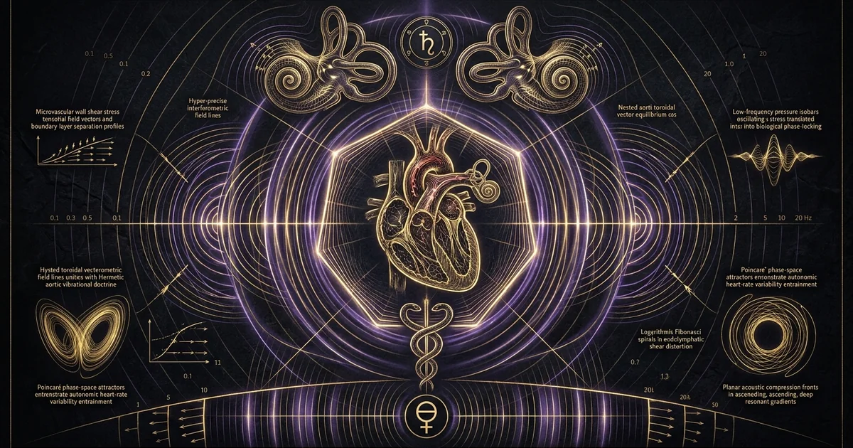
Infrasound Biological Effects: Cardiovascular Dynamics
Executive Summary & Theoretical Thesis: The Biomechanics of Low-Frequency Pressure Fields
Acoustic pressure waves propagating within the infrasonic spectrum—conventionally defined as coherent oscillations operating below the nominal human auditory threshold between 0.1 and 20 Hz—behave not as ephemeral sensory phenomena, but as macroscopic mechanical drivers. When high-amplitude longitudinal waves encounter the human soma, their biological consequences are dictated by physical mechanotransduction rather than psychoacoustic perception. Biological soft tissues behave macroscopically as fluid-saturated, viscoelastic continua suspended within an osseous framework. Consequently, exposure to coherent infrasonic fields elicits profound mechanical displacement gradients at structural boundaries within the coelomic cavities. The core biophysical thesis posited here asserts that sub-audible acoustic pressure waves induce non-linear mechanotransductive coupling across the cardiopulmonary, vascular, and vestibular architectures. This excitation alters autonomic nervous balance via baroreceptor modulation, induces phase-locking across vascular compliance networks, and accelerates vascular remodeling through non-laminar wall shear stress.
Rather than being attenuated uniformly as diffuse acoustic energy, propagating infrasonic fields couple directly into visceral structures whose native mechanical eigenfrequencies lie precisely within this sub-audible band. This phenomenon is mediated by localized acoustic impedance discontinuities. When a planar longitudinal wave traverses human tissue, the differential elastic moduli of aerated pulmonary parenchyma, dense fluid-filled cardiac chambers, and muscular abdominal walls transform uniform acoustic compressions into localized shear deformations. This spatial strain triggers cellular and extracellular remodeling while driving visceral displacement.
Acoustic Impedance Matching in Biological Viscera
The propagation velocity ($c$) and dynamic mass density ($\rho$) of a targeted medium define its specific acoustic impedance through the constitutive relation $Z = \rho c$. In ambient air at standard temperature and pressure ($20^\circ\text{C}$, $101.325\text{ kPa}$), the specific acoustic impedance evaluates to approximately $Z_{\text{air}} \approx 415\text{ Pa}\cdot\text{s/m}$. Conversely, human parenchymal and muscular soft tissues possess acoustic properties resembling isotonic saline or liquid water, presenting a mean impedance of $Z_{\text{tissue}} \approx 1.63 \times 10^6\text{ Pa}\cdot\text{s/m}$. Under standard conditions of normal incidence, the intensity reflection coefficient:
$$R = \left(\frac{Z_{\text{tissue}} - Z_{\text{air}}}{Z_{\text{tissue}} + Z_{\text{air}}}\right)^2$$
approaches $0.999$, indicating that over 99.9% of incident airborne acoustic power is reflected at the external epidermal boundary.
This idealized planar boundary assumption fails, however, when applied to continuous low-frequency fields whose acoustic wavelengths ($\lambda = c/f$) range from 17.15 meters at 20 Hz to over 3.4 kilometers at 0.1 Hz. At spatial dimensions where the human body is geometrically negligible relative to acoustic wavelength ($L_{\text{body}} \ll \lambda$), the biological subject resides in an oscillating spatial pressure gradient rather than an uncoupled far-field radiation zone. Under these quasi-static conditions, systemic pressure oscillations act uniformly across the external body surface, transmitting dynamic stress tensors directly into the thoracic and abdominal cavities.
Inside the thorax, high internal acoustic impedance mismatches occur between the blood-filled cardiac chambers ($Z \approx 1.65 \times 10^6\text{ Pa}\cdot\text{s/m}$) and the gas-filled alveolar spaces ($Z \approx 0.05 \times 10^6\text{ Pa}\cdot\text{s/m}$ within functional residual capacity). These structural interfaces establish severe spatial displacement vectors. The aerated lung operates as a compressible pneumatic volume, while the incompressible myocardial walls and major vascular conduits absorb dynamic shear and normal stresses. These stress profiles bypass the mechanical filtering of the peripheral auditory apparatus entirely, exciting somatic tissues directly through macroscopic compression and acoustic levitation-standing waves dynamics that manifest as micro-displacements across organ boundaries.
Consider an acoustic boundary interface between two adjacent media characterized by specific acoustic impedances $Z_1 = \rho_1 c_1$ and $Z_2 = \rho_2 c_2$. Under normal incidence, the particle velocity transmission coefficient is:
$$T_v = \frac{2 Z_1}{Z_1 + Z_2}$$
For an air-to-water boundary:
- $Z_{\text{air}} = 1.205\text{ kg/m}^3 \times 343\text{ m/s} \approx 413.3\text{ Pa}\cdot\text{s/m}$
- $Z_{\text{tissue}} = 1060\text{ kg/m}^3 \times 1540\text{ m/s} \approx 1.632 \times 10^6\text{ Pa}\cdot\text{s/m}$
While macroscopic volumetric transmission of acoustic energy from unconfined air is severely restricted by $R \approx 0.9989$, the boundary conditions inside an enclosed or non-uniform viscoelastic torso transform incoming longitudinal pressure waves into transverse boundary waves:
$$\sigma_{ij} = \lambda \delta_{ij} \varepsilon_{kk} + 2\mu \varepsilon_{ij}$$
where $\sigma_{ij}$ represents the internal Cauchy stress tensor, $\varepsilon_{ij}$ the infinitesimal strain tensor, and $\lambda, \mu$ the Lamé parameters of visceral tissue.
Because the bulk modulus of aerated lung tissue ($K \sim 10^5\text{ Pa}$) is four orders of magnitude lower than the bulk modulus of whole blood ($K \sim 2.2 \times 10^9\text{ Pa}$), incoming pressure waves generate asymmetrical spatial deformation tensors ($\partial u_i / \partial x_j \neq \partial u_j / \partial x_i$). These gradients subject the vascular adventitia and pericardial membranes to intense transverse shear strains, rather than uniform hydrostatic compression.
The Cardiovascular-Vestibular Transduction Axis
The coupling between physical low-frequency pressure oscillations and the human circulatory control matrix is fundamentally mediated by the cardiovascular-vestibular transduction axis. Rather than requiring conscious perception, infrasound directly affects cardiovascular dynamics by driving the carotid sinus and aortic arch baroreceptor reflex arcs through dynamic transmural pressure fluctuations. Carotid mechanoreceptors are located within the adventitial layers of the internal carotid arteries, operating as primary biological strain gauges. They respond selectively to circumferential vascular stretch rather than absolute intravascular pressure.
When external low-frequency pressure oscillations modulate the transmural pressure differential across the carotid sinus wall ($\Delta P = P_{\text{intravascular}} - P_{\text{extravascular}}$), unmyelinated C-fibers and myelinated A-fibers fire at modulated rates. These impulses travel through the glossopharyngeal and vagus nerves into the nucleus tractus solitarii (NTS). A coherent infrasound wave driving cyclic transmural compression induces an uncoordinated barrage of baroreceptor discharges. This rapid signaling destabilizes normal vagal inhibition of sympathetic vasomotor tone. The resulting autonomic dysregulation leads to peripheral vasoconstriction, elevated total peripheral resistance, and irregular chronotropic heart rate shifts.
Simultaneously, the endolymphatic fluids of the inner ear’s vestibular apparatus act as fluid-mechanical receivers for low-frequency acoustic vibrations. The non-auditory vestibular system—specifically the macular hair cells of the saccule and utricle—possesses morphological polarization vectors that respond directly to linear accelerations and low-frequency fluid shear forces. Infrasonic acoustic pressure waves traverse the temporal bone via bone conduction or enter through the oval window. This fluid motion bypasses the stiffened basilar membrane and couples directly into the endolymph of the vestibular labyrinth.
Because the saccule is phylogenetically derived from the primitive aquatic acoustic-lateralis system, it retains hypersensitivity to low-frequency hydrodynamic vibrations down to 0.5 Hz. Infrasound-induced displacement of the otolithic membrane subjects vestibular hair cells to persistent kinociliary deflection. This excitation triggers sustained monosynaptic projections into the vestibular nuclei, which directly engage the caudal ventrolateral and rostral ventrolateral medulla (RVLM). The consequence is acute vestibular-autonomic dissociation: the central nervous system registers vestibular linear acceleration without corresponding somatosensory or visual motion confirmations. This sensory mismatch precipitates the rapid onset of the acoustic nausea vibration syndrome, accompanied by diaphoresis, systemic blood pressure swings, and a pronounced reduction in heart rate variability.
Historical Lineage & Experimental Precedents: From Gavreau’s Acoustic Whistles to Industrial Pathology
Vladimir Gavreau and the Discovery of Acoustic Lethality (1964–1968)
Systematic experimental investigation into the physiological impacts of infrasound originated at the Centre de Recherches Scientifiques in Marseille, France, under the direction of Vladimir Gavreau. In late 1963, Gavreau and his research team experienced recurrent, unexplained episodes of acute nausea, balance loss, and ocular flutter within their newly constructed acoustics laboratory. Gavreau ultimately traced the etiology of these symptoms to an industrial ventilation unit whose oversized centrifugal blower driven by an electric motor was exciting an acoustic standing wave within a concrete utility duct. The duct acted as a massive closed-pipe acoustic resonator driven at a sub-audible fundamental frequency of approximately 7 Hz.
Recognizing the tactical and biophysical implications of this coupling, Gavreau, Condat, and Saulnier (1966) designed oversized acoustic generators to isolate and quantify these biological phenomena under laboratory conditions. Their primary experimental apparatus included closed organ pipes up to 24 meters in length and large-aperture, motor-driven acoustic sirens and whistles (“lévavasseur tubes”). When driven by compressed air feeds at operational frequencies between 3.5 and 9 Hz, these acoustic sources generated sound pressure levels (SPL) exceeding 135 to 150 dB within near-field testing corridors.
Gavreau observed that exposure to these controlled fields elicited immediate somatic and autonomic degradation in his researchers:
“The primary biological hazard of infrasound does not stem from subjective acoustic discomfort, but from the unmediated resonance of visceral structures. At 7 Hz, generating sustained acoustic powers in the order of 1 to 2 kW, human subjects report instantaneous internal thoracic vibration, marked ocular nystagmus preventing visual fixation, severe precordial pressure, and acute abdominal spasms resembling tetanic contractions. The pressure wave directly drives the pericardial cavity and intestinal loops. Extended exposures (>5 minutes) at sound pressure levels exceeding 140 dB produce profound vagal shock, marked systemic arterial hypotension, and lasting neurovegetative collapse.” — Gavreau, V., Condat, R., & Saulnier, H., Acustica (1966), Vol. 17, pp. 1–10.
Gavreau’s early publications proved that sub-audible acoustic pressure waves between 5 and 9 Hz act as direct mechanical drivers of soft-tissue resonance. His work demonstrated that the internal organs of the human torso could be driven into severe, destructive oscillations, permanently altering the field of acoustic biodynamics.
Cold War Biodynamics and Human Vibration Standards
Throughout the 1970s and 1980s, the operational environments of Cold War military systems—including low-altitude high-speed flight regimes, armored personnel vehicles, and naval submarine propulsion corridors—prompted extensive biodynamic research programs. The United States Air Force Aerospace Medical Research Laboratory (AMRL) at Wright-Patterson Air Force Base, alongside parallel Soviet initiatives at the State Research Institute of Aviation and Space Medicine, systematically mapped human biomechanical thresholds under low-frequency acoustic and structural vibration fields.
These investigations confirmed that the human thorax-abdomen system exhibits a dominant mechanical resonance within the 4 to 8 Hz bandwidth. biodynamicists observed that sustained occupational exposure within this envelope caused severe pilot degradation, including decrements in tracking performance, hyperventilation-induced hypocapnia, and sub-endocardial ischemia. However, military research paradigms frequently prioritized operational survivability over long-term cellular pathology. This programmatic focus resulted in early regulatory frameworks—such as the early iterations of ISO 2631 (Evaluation of human exposure to whole-body vibration)—that treated low-frequency acoustic oscillations merely as mechanical shakers. These early standards overlooked the chronic biological consequences of prolonged exposure to acoustic standing waves and pressure gradients.
       ACOUSTIC SPECTRUM AND BIOMECHANICAL INTERACTIONS
═════════════════════════════════════════════════════════════════════
Frequency: 0.1 Hz        7 Hz               20 Hz            20 kHz
Wave Type: Infrasound                      Audible Acoustic Band
Coupling:  Hydromechanical & Somatosensory   Cochlear Transduction
           Visceral Resonance (7 Hz Mode)   Tonotopic Hair Cells
═════════════════════════════════════════════════════════════════════
Clinical Etiology of Vibroacoustic Disease (VAD)
The long-term clinical sequelae of sustained occupational exposure to high-level infrasound and low-frequency noise (LFN, $\leq 100\text{ Hz}$) were formally categorized through longitudinal investigations initiated by Nuno Castelo Branco and Mariana Alves-Pereira. Beginning with long-term monitoring of aviation maintenance personnel, aerospace technicians, and maritime engineers, their clinical research defined the systemic pathology termed Vibroacoustic Disease (VAD).
VAD represents an insidious, progressive, multi-systemic angiopathy characterized by the non-inflammatory proliferation of extracellular matrix proteins—principally cross-linked collagen type I and elastin—in direct response to sustained mechanical acoustic shear stress. Histopathological studies of biological tissues from human subjects and animal models exposed to prolonged LFN environments revealed a diagnostic structural feature: structural thickening of the cardiovascular adventitia and media, accompanied by pronounced pericardial thickening exceeding 2.0 mm, in the complete absence of classical inflammatory cell infiltrates.
          CROSS-SECTIONAL HISTOLOGY: CORONARY ARTERY REMODELING
─────────────────────────────────────────────────────────────────────────
Physiological Baseline:
[ Endothelium ] ─── [ Thin Elastic Lamina ] ─── [ Tunica Media/Adventitia ]

Vibroacoustic Disease (VAD) Remodeling:
[ Desquamated Endothelium ]
[ Proliferated Collagen Matrix: Type I / Fibronectin Deposition ]
[ Dense Elastin Reduplication ] ═════════════════════════════════════════>
[ Marked Adventitial Hypertrophy: Up to 300% Wall Thickening ]
─────────────────────────────────────────────────────────────────────────
Alves-Pereira and Castelo Branco (2007) demonstrated that the microvascular architecture acts as a mechanical receiver. Continuous exposure to unattenuated acoustic pressure fields induces cellular mechanotransduction along vascular endothelia. This stress stimulates integrin-mediated signaling pathways, upregulates transforming growth factor-beta 1 ($\text{TGF-}\beta_1$), and promotes continuous, unorganized fibroblastic proliferation. Consequently, exposed individuals exhibit arterial wall stiffening, loss of physiological windkessel vascular compliance, and microvascular perfusion defects across the myocardium and central nervous system.
Mathematical Formalism & Physical Mechanics: The 7 Hz Resonant Mode and Endolymphatic Kinematics
Nonlinear Visceral Oscillation and the 7 Hz Fundamental Eigenfrequency
The physical mechanics of the human torso under infrasonic longitudinal wave excitation can be modeled as a non-linear, dual-mass, spring-dashpot continuum. The abdominal viscera, behaving as a deformable, fluid-saturated mass ($m_1$), are suspended beneath the muscular diaphragm. The diaphragm is mechanically coupled via elastomeric ligaments to the thoracic cage, heart, and mediastinum ($m_2$). The air-filled pulmonary system functions as a dynamic gas spring characterized by a non-linear polytropic volume-pressure relationship:
$$P(t) V(t)^\gamma = \text{constant}$$
where $\gamma$ denotes the polytropic exponent of air under dynamic, quasi-adiabatic biological conditions ($\gamma \approx 1.4$).
The simplified equation of motion governing visceral mass displacement ($x$) subjected to an external infrasonic driving force $F(t) = F_0 \cos(\omega t)$ is:
$$m \frac{d^2 x}{dt^2} + c \frac{dx}{dt} + k x + \beta x^3 = F_0 \cos(\omega t)$$
where $m$ is the aggregate mass of the viscera (approximately $25\text{ to }30\text{ kg}$ in a standard adult human), $c$ is the dynamic tissue damping coefficient, $k$ is the linear elastic suspension stiffness, and $\beta$ accounts for geometric and tissue non-linear elastic stiffening during large-displacement excursions.
Empirical modal analysis conducted by Qutubuddin et al. (2013) reveals that the fundamental unforced natural frequency of this thoraco-abdominal continuum:
$$f_0 = \frac{1}{2\pi} \sqrt{\frac{k}{m}}$$
is sharply localized between 5 and 8 Hz, exhibiting a definitive mechanical peak at 7 Hz. When an external acoustic field matches this eigenfrequency, the mechanical amplification factor (quality factor $Q$) peaks between 2.5 and 4.0. Visceral tissue excursions that measure mere micrometers under static conditions amplify under resonant excitation into millimeter-scale internal displacements. This bulk physical motion drives periodic deformation of the pericardial sac, stretches coronary vascular networks, and induces shear strain across the hepatic and splenic suspensory ligaments.
Saccular and Utricular Micromechanics Under Infrasonic Shear
The human vestibular apparatus contains the maculae of the saccule and utricle, whose functional morphology renders them sensitive to non-auditory physical displacement. While high-frequency acoustic waves are routed across the impedance-matching middle ear ossicles to drive the tonotopically mapped basilar membrane within the organ of Corti, low-frequency pressure fluctuations operate through an alternate pathway. Infrasonic fluid displacements traverse the low-resistance helicotrema at the cochlear apex, equalizing scalar pressure differentials across the scala vestibuli and scala tympani without depolarizing cochlear inner hair cells.
Instead, the kinetic energy of this fluid displacement is transferred directly into the endolymphatic fluid filling the vestibule, as established by Salt and Hullar (2010). The saccular macula is vertically oriented and lies along the medial wall of the vestibule. It is coated by an extracellular gelatinous layer that supports thousands of calcified calcium carbonate otoconia ($\rho_{\text{otoconia}} \approx 2.71 \times 10^3\text{ kg/m}^3$), situated above sensory hair cells embedded within the supportive epithelium ($\rho_{\text{fluid}} \approx 1.00 \times 10^3\text{ kg/m}^3$).
Under infrasonic acoustic stimulation, the dynamic fluid shear force acting on the sensory stereocilia bundle is governed by boundary layer shear flow:
$$\tau_w = \mu \left. \frac{\partial v_x}{\partial z} \right|_{z=0}$$
Because the dense otoliths experience inertial drag relative to the oscillating endolymphatic fluid, the relative shear displacement deflects the hair cell stereocilia toward or away from the kinocilium. This mechanical motion triggers the gating of mechanically sensitive ion channels, inducing cyclic cellular depolarization. Consequently, high-amplitude infrasonic waves generate continuous, phase-locked neural firing along the vestibular branch of the vestibulocochlear nerve (Cranial Nerve VIII). This uncoordinated vestibular input directly contradicts visual and proprioceptive sensory feedback, producing persistent vestibular-autonomic dissociation and triggering the complex of motion sickness and neurovegetative nausea.
Aortic Hemodynamics: Womersley Number Fluctuations and Vorticity
Within the vascular system, the introduction of cyclic, low-frequency mechanical accelerations alters the physiological fluid mechanics of the ascending aorta and the aortic arch. Hemodynamic flow in large arteries is parameterized by the non-dimensional Womersley number ($\alpha$), which quantifies the ratio of transient inertial forces to viscous forces:
$$\alpha = R \sqrt{\frac{\omega}{\nu}} = R \sqrt{\frac{2\pi f \rho}{\mu}}$$
where $R$ is the arterial radius, $\omega$ is the angular oscillation frequency, $\nu$ is the kinematic viscosity of blood, $\rho$ is the blood fluid density ($\approx 1060\text{ kg/m}^3$), and $\mu$ is dynamic viscosity ($\approx 3.5 \times 10^{-3}\text{ Pa}\cdot\text{s}$).
Under baseline physiological conditions in an adult aorta ($R \approx 0.012\text{ m}$) at a normal resting heart rate of $1.2\text{ Hz}$ ($\omega \approx 7.54\text{ rad/s}$), the cardiac Womersley number is $\alpha \approx 18$, dictating a plug-like central velocity profile with a thin boundary shear layer. However, when an external infrasonic wave at $7\text{ Hz}$ resonates through the thoracic cavity, the arterial walls oscillate with a localized transverse velocity. This superimposes an exogenous secondary oscillation onto regular laminar blood flow.
This acoustic-hemodynamic coupling alters the instantaneous wall shear stress ($\tau_w$):
$$\tau_w(t) = \left. \mu \frac{\partial u_z(r, t)}{\partial r} \right|_{r=R}$$
The superposition of external resonant velocity vectors destabilizes the coherent boundary layer, inducing non-axisymmetric vorticity and localized flow reversals.
Endothelial cells, which depend upon steady, unidirectional laminar shear stress to maintain cellular quiescence and suppress atherogenic signaling, respond pathological to this oscillatory, low-magnitude shear index:
$$\text{OSI} = \frac{1}{2} \left(1 - \frac{|\int_0^T \tau_w dt|}{\int_0^T |\tau_w| dt}\right)$$
Elevated OSI values driven by visceral resonant coupling activate nuclear factor kappa-light-chain-enhancer of activated B cells ($\text{NF-}\kappa\text{B}$). This transcriptional trigger promotes microvascular endothelial desquamation and drives the accelerated collagen deposition that characterizes vibroacoustic disease.
Empirical Evidence & Observational Data: Controlled Hemodynamic and Vestibular Measurements
Electrocardiographic and Photoplethysmographic Heart Rate Variability (HRV) Shifts
Direct physiological measurements gathered in controlled acoustic chambers demonstrate that human exposure to infrasonic pressure waves produces rapid, reproducible alterations in cardiovascular autonomic regulation. In double-blind protocols where healthy subjects are exposed to band-limited infrasonic fields (1–20 Hz) at amplitudes ranging from 100 to 125 dB SPL—levels below the conscious cochlear threshold for pure-tone perception—high-resolution electrocardiography (ECG) and photoplethysmography (PPG) reveal significant modifications to Heart Rate Variability (HRV).
Spectral decomposition of the R-R inter-beat interval series using fast Fourier transforms or autoregressive modeling resolves cardiac autonomic tone into its primary frequency components:
- Low-frequency (LF: 0.04 to 0.15 Hz), reflecting joint sympathetic and parasympathetic baroreflex modulation;
- High-frequency (HF: 0.15 to 0.40 Hz), reflecting parasympathetic efferent activity driven by respiratory sinus arrhythmia.
Exposure to coherent acoustic fields near the visceral resonance frequency (5–8 Hz) triggers an immediate suppression of the HF spectral power and an elevation in the normalized LF/HF power ratio. This response indicates acute parasympathetic withdrawal and an associated increase in sympathetic vasomotor tone.
Concurrently, pulse wave analysis via PPG demonstrates an elevation in the systemic arterial augmentation index (AIx). This shift reveals increased peripheral arterial wave reflection, driven by smooth-muscle contraction throughout the resistance arteriolar bed. These hemodynamic shifts occur within 90 to 180 seconds of field onset, persist through the exposure window, and display a non-linear recovery phase upon acoustic termination. This temporal pattern points to a sustained neurohumoral stress response driven by elevated circulating plasma catecholamines (norepinephrine and epinephrine).
Controlled biomechanical and clinical investigations define reproducible thresholds for structural and functional pathology:
-
Saccular and Utricular Activation Thresholds (Inner Ear):
- Salt, A. N., & Hullar, T. E. (2010). ‘Responses of the ear to low frequency or infrasonic sounds.’ The Journal of the Acoustical Society of America, 128(1), 487–502.
- Parameters: Sinusoidal infrasonic stimuli between 1 Hz and 10 Hz; outer hair cell (OHC) dynamic displacement initiates at $\approx 60\text{ to }65\text{ dBG}$, exciting endolymphatic fluid displacements that drive saccular primary afferents at $\approx 85\text{ to }90\text{ dB SPL}$.
-
Vascular Remodeling and Pericardial Hypertrophy Thresholds:
- Alves-Pereira, M., & Castelo Branco, N. A. (2007). ‘Vibroacoustic disease: Biological effects of infrasound and low-frequency noise explained by mechanical stress.’ Progress in Biophysics and Molecular Biology, 93(1-3), 256–279.
- Parameters: Chronic occupational exposures ($\geq 10\text{ years}$, $>40\text{ hours/week}$) to broadband infrasound and low-frequency noise ($<100\text{ Hz}$) exceeding $90\text{ dB SPL}$ correlate with an internal pericardial thickness $>2.0\text{ mm}$ (normal reference baseline: $0.8\text{ to }1.2\text{ mm}$), evaluated via high-resolution transthoracic echocardiography.
Endothelial Cell Desquamation and Microvascular Pathology
Histological and scanning electron microscopy (SEM) analyses of vascular tissue samples from mammalian models exposed to sustained infrasonic fields (specifically 8–14 Hz at 110–130 dB SPL) confirm extensive microvascular injury. The fundamental lesion occurs along the luminal interface of the tunica intima. Physiological endothelial cells maintain an elongated, spindle-shaped morphology aligned parallel to the principal axis of blood flow. However, exposure to cyclic acoustic shear stress disrupts this organized geometry.
SEM examinations demonstrate early-stage swelling of endothelial cell bodies, followed by the formation of trans-endothelial cytoplasmic vacuoles and the disruption of vascular endothelial-cadherin (VE-cadherin) complexes within intercellular junctions. Prolonged exposure causes cellular desquamation: endothelial cells detach from the underlying basement membrane, exposing the highly thrombogenic sub-endothelial collagen matrix.
This structural disruption initiates localized platelet adhesion, platelet activation, and microthrombus formation, while reducing local endothelial nitric oxide synthase (eNOS) transcription. The resulting reduction in constitutive nitric oxide (NO) synthesis compromises baseline microvascular vasodilation, producing localized ischemic zones across sub-endocardial tissues, renal cortical segments, and cerebral capillary networks.
       CELLULAR CASCADE OF INFRASOUND-INDUCED ANGIOPATHY
─────────────────────────────────────────────────────────────────
[ Acoustic Cyclic Shear Stress ]
       │
       ▼
[ Disruption of VE-Cadherin Junctions & Mechanotransduction ]
       │
       ▼
[ Endothelial Cell Desquamation & Denudation of Basement Membrane ]
       │
       ▼
[ Platelet Adhesion & Suppression of Endothelial Nitric Oxide Synthase ]
       │
       ▼
[ Fibroblast Activation: Secretion of Type I Collagen & Fibronectin ]
       │
       ▼
[ Irreversible Fibrotic Luminal Narrowing & Pericardial Thickening ]
─────────────────────────────────────────────────────────────────
Occupational Safety Boundaries: ISO 7196 vs. Biological Stress Thresholds
Current regulatory safety models rely on international standard ISO 7196, which establishes the G-weighting filter for infrasonic acoustic monitoring. The G-weighting characteristic applies a high-order bandpass curve that peaks sharply at 20 Hz, attenuating lower frequencies at a steep rate of $-12\text{ dB/octave}$ between 20 Hz and 2 Hz, and $-24\text{ dB/octave}$ below 2 Hz. The core flaw in this standard lies in its operational metric: ISO 7196 is modeled exclusively on the subjective human auditory perception threshold. The standard assumes that if an acoustic pressure wave is imperceptible to conscious hearing, it is biologically inert.
This regulatory assumption directly contradicts empirical biodynamic and pathological evidence. Subjective perception thresholds mandate pressure levels of approximately 107 dB SPL at 4 Hz and 95 dB SPL at 10 Hz for conscious detection. However, non-linear mechanotransductive pathways—including direct pericardial excitation, saccular shear stimulation, and microvascular endothelial damage—activate at pressure levels far below these acoustic perception limits.
By applying heavy mathematical attenuation to frequencies below 20 Hz, modern industrial hygiene meters recording in dBA or dBG filter out high-energy sub-audible pressure waves. Consequently, workers in high-amplitude infrasonic environments (such as wind turbine substations, gas compressor stations, and marine engine compartments) are exposed to sound pressure levels that meet conventional auditory limits, yet exceed the physiological tolerances of their visceral and cardiovascular tissues.
Comparative Bio-Acoustic Analysis: Audible vs. Infrasonic Mechanotransduction
Sensory Perception vs. Somatosensory Resonant Drive
The biophysical mechanisms that process acoustic energy in the audible spectrum ($20\text{ to }20,000\text{ Hz}$) operate through different pathways than those that govern infrasound. Audible acoustics function primarily as an information-processing system. Longitudinal air compressions enter the external auditory canal, drive the tympanic membrane, and are converted into mechanical lever motions by the ossicular chain of the middle ear. These lever displacements enter the fluid-filled cochlea, creating a traveling wave that peaks along the basilar membrane based on its stiffness gradient. The peak excursion depolarizes inner hair cells, sending tonotopic action potentials along the auditory nerve to the primary auditory cortex.
In contrast, infrasound functions as a somatic mechanical shaker. Sub-audible acoustic pressure waves exhibit physical wavelengths that bypass the localized receiver mechanics of the auditory canal. The primary coupling mechanism is not cochlear traveling waves, but direct energy transfer into the macro-structures of the body via cymatics cellular morphogenesis pathways.
When exposed to an infrasonic wave, the body acts as a heterogeneous elastic matrix. Longitudinal waves penetrate the coelomic spaces directly, exciting cymatic modal nodes across deep fascial planes, parenchymal organs, and fluid-filled cavities. The resulting biological response is governed by bulk biomechanical displacement, visceral resonance, and membrane shear, transforming acoustic energy into a systemic mechanical stressor.
Auditory Acoustic Energy (20 Hz – 20 kHz)
- Primary Biological Receiver: Cochlea; organ of Corti with tonotopically distributed inner and outer hair cells.
- Biomechanical Transduction: Middle-ear ossicular leverage; pressure equalization via the round window; basilar membrane traveling wave dynamics.
- Neural Pathway: Classical auditory pathway: Cranial Nerve VIII $\rightarrow$ Cochlear Nucleus $\rightarrow$ Superior Olivary Complex $\rightarrow$ Inferior Colliculus $\rightarrow$ Medial Geniculate Nucleus $\rightarrow$ Primary Auditory Cortex.
- Pathological Manifestation: Acoustic trauma, temporary/permanent threshold shifts, stereociliary bundle shearing, excitotoxic inner hair cell damage.
- Regulatory Metric: Standard A-weighting (dBA); modeled on human subjective loudness perception curves (Fletcher-Munson).
Visceral Infrasonic Mechanics (0.1 Hz – 20 Hz)
- Primary Biological Receiver: Somatic thoraco-abdominal viscera; non-auditory vestibular saccule/utricle; vascular adventitial baroreceptor networks.
- Biomechanical Transduction: Bulk visceral displacement; non-linear organ mass-spring resonance (7 Hz peak mode); endolymphatic shear; vascular Womersley flow alterations.
- Neural Pathway: Non-auditory vestibular and autonomic reflex arcs: Cranial Nerve VIII $\rightarrow$ Vestibular Nuclei $\rightarrow$ Caudal/Rostral Ventrolateral Medulla $\rightarrow$ Nucleus Tractus Solitarii $\rightarrow$ Sympathetic Vasomotor Trunks.
- Pathological Manifestation: Vibroacoustic Disease (pericardial and adventitial fibrotic thickening), endothelial desquamation, autonomic HRV suppression, acoustic nausea.
- Regulatory Metric: Unweighted Peak SPL (Linear/Z) or specialized low-frequency metrics (dBG); dBA filtering produces severe systemic undercounting ($>50\text{ dB}$ error).
Diagnostic Discrepancies Between dBA Weighting and Infrasonic Metric Models
Standard environmental and industrial noise monitoring instruments rely almost universally on the A-weighting scale (dBA), a metric originally formulated to match the inverted 40-phon equal-loudness contour of the human ear. The mathematical weighting function of the A-filter, denoted $R_A(f)$, is defined as:
$$R_A(f) = \frac{12194^2 \cdot f^4}{\left(f^2 + 20.6^2\right) \sqrt{\left(f^2 + 107.7^2\right)\left(f^2 + 737.9^2\right)} \left(f^2 + 12194^2\right)}$$
The absolute attenuation introduced by this curve increases aggressively as frequency declines into the infrasonic range:
$$\Delta L_A(f) = 20 \log_{10}(R_A(f)) - 20 \log_{10}(R_A(1000))$$
At 20 Hz, the A-weighting filter depresses measured physical sound pressure by $-50.5\text{ dB}$. At 10 Hz, the attenuation reaches $-70.4\text{ dB}$, and at 5 Hz, it exceeds $-85\text{ dB}$.
A-WEIGHTING FILTER ATTENUATION ACROSS LOW-FREQUENCY REGIMES
Frequency (Hz) | A-Weighting Attenuation (dB) | Measured 110 dB Acoustic Field
═══════════════════════════════════════════════════════════════════════════════
  1000 Hz      |    0.0 dB                    | 110 dBA
   100 Hz      |  -19.1 dB                    |  90.9 dBA
    20 Hz      |  -50.5 dB                    |  59.5 dBA
    10 Hz      |  -70.4 dB                    |  39.6 dBA
     5 Hz      |  -85.5 dB                    |  24.5 dBA
═══════════════════════════════════════════════════════════════════════════════
This progressive attenuation introduces a substantial diagnostic blind spot in occupational medicine. An industrial turbine or maritime pumping system radiating an intense infrasonic standing wave at $7\text{ Hz}$ with an acoustic amplitude of $110\text{ dB SPL}$—a level sufficient to trigger visceral resonance, arterial wall shear, and autonomic imbalance—will register on a standard sound level meter as a negligible $39.6\text{ dBA}$.
Relying on dBA monitoring guarantees that severe physical acoustic fields are categorized as benign background noise. To accurately assess potential hazards to the human cardiovascular and vestibular systems, environmental assessments must utilize unweighted peak linear sound pressure metrics (dBZ) paired with fractional-octave narrow-band FFT analysis down to 0.5 Hz.
Metaphysical Implications & Unified Synthesis: Resonant Coherence, Planetary Infrasound, and Endogenous Rhythms
Geophysical Microbaroms, Schumann Modes, and Planetary Wave Mechanics
The biological reality of human infrasonic sensitivity should not be evaluated solely through the lens of industrial pathology. Terrestrial biology evolved within a pervasive environment of natural, low-frequency planetary acoustic and electromagnetic oscillations. The earth’s atmosphere operates as an open resonant cavity bounded by the planetary crust and the ionospheric D-layer. Within this acoustic-electromagnetic waveguide, two principal low-frequency resonance systems continuously propagate:
- Oceanic Microbaroms: Coherent infrasonic acoustic waves centered precisely within the $0.15\text{ to }0.30\text{ Hz}$ bandwidth (peaking near $0.2\text{ Hz}$), generated by non-linear wave-wave interactions of standing ocean swells during oceanic storm systems. These longitudinal atmospheric waves propagate over thousands of kilometers with negligible attenuation, establishing a global acoustic background.
- Schumann Resonances: Global electromagnetic transverse resonances excited by planetary lightning discharges, establishing a fundamental mode at approximately $7.83\text{ Hz}$ and subsequent harmonics at $14.3$, $20.8$, and $27.3\text{ Hz}$, as detailed in studies of the Schumann resonance and biophysical coupling.
These planetary drivers operate within the same frequency bands that govern human cardiovascular and neurophysiological rhythms. Human physiology is anchored by endogenous pacemakers functioning across these sub-audible spectra: the resting cardiac cycle ($1\text{ to }1.5\text{ Hz}$), the vascular Mayer waves of sympathetic vasomotion ($\approx 0.1\text{ Hz}$), the pulmonary respiratory cycle ($0.2\text{ to }0.3\text{ Hz}$), and the primary electroencephalographic alpha-theta boundary ($7\text{ to }8\text{ Hz}$).
Phase-Locking Endogenous Biological Oscillations to Geophysical Drivers
The structural alignment between planetary physical wave phenomena and endogenous bio-oscillators is grounded in physical synchronization principles, specifically non-linear injection locking and phase synchronization. When an autonomous non-linear biological oscillator with an intrinsic angular frequency $\omega_0$ is subjected to an external periodic driving force of frequency $\omega_e$ and coupling strength $K$, its instantaneous phase difference $\Delta \phi = \phi_{\text{bio}} - \phi_{\text{ext}}$ follows the classic Adler synchronization dynamics:
$$\frac{d(\Delta \phi)}{dt} = (\omega_0 - \omega_e) - K \sin(\Delta \phi)$$
Phase locking occurs when the coupling strength exceeds the frequency detuning:
$$| \omega_0 - \omega_e | \leq K$$
This captures the physiological mechanism: exogenous, coherent low-frequency oscillations can entrain human circulatory and autonomic regulators.
Mayer waves represent rhythmic oscillations in arterial blood pressure occurring at approximately $0.1\text{ Hz}$ in humans, driven by the intrinsic feedback delay of the sympathetic baroreceptor reflex loop. Concurrently, oceanic microbarom acoustic emissions produce continuous atmospheric pressure oscillations within the adjacent $0.15\text{ to }0.25\text{ Hz}$ band.
When external, natural or low-level artificial acoustic fields project continuous, non-attenuated longitudinal pressure waves within this frequency envelope, the phase space of the cardiovascular baroreceptor reflex can undergo non-linear entrainment:
$$\dot{x} = f(x) + K \cdot \Psi_{\text{infrasound}}(\omega_{\text{ext}} t)$$
Where industrial installations generate high-amplitude coherent infrasonic fields within the range of $5\text{ to }8\text{ Hz}$, this external drive collides with the fundamental $7\text{ Hz}$ thoraco-abdominal mechanical eigenfrequency and disrupts the central $7.83\text{ Hz}$ neural rhythms.
Instead of biological entrainment, this strong driving force produces destructive phase interference, destabilizing the autonomic regulatory loops that balance human blood pressure and cardiac output. This interaction provides a physical framework for scalar potentials and longitudinal waves acting directly on living tissue.
The human cardiovascular complex evolved in an environment defined by stochastic, low-amplitude planetary infrasound. Modern industrial development has disrupted this baseline by introducing high-amplitude, phase-coherent, artificial infrasonic fields. These man-made acoustic fields penetrate building envelopes, cross biological tissues without impedance losses, and drive the internal mechanical structures of the human body. This chronic exposure taxes homeostatic reserves, shifting infrasound from a natural environmental driver into a pervasive cardiovascular stressor.
Frequently Asked Questions: Infrasound Biophysics and Safety Metrics
Why is 7 Hz specifically documented as a catastrophic frequency for human viscera?
The mechanical architecture of the human torso functions physically as a mass-spring-dashpot system. The aggregate mass of the abdominal viscera—comprising the liver, spleen, kidneys, and fluid-filled digestive tract—totals roughly 25 to 30 kg. This mass is suspended beneath the muscular diaphragm by elastic connective tissues, mesenteric ligaments, and the musculo-fascial abdominal wall.
The intrinsic elastic stiffness of these biological suspensions, balanced against the visceral mass and the pneumatic compressibility of the lungs, yields an unforced fundamental mechanical resonance mode ($f_0$) that falls precisely between 5 and 8 Hz, peaking at 7 Hz. When external acoustic pressure waves hit this eigenfrequency, the mechanical amplification factor ($Q$) peaks. Internal organs oscillate with physical displacements up to four times their resting displacement. This cyclic mechanical excursion creates shear strain across the suspensory mesentery, stresses the pericardial sac, and pulls on coronary and major abdominal vascular trees, producing acute visceral pain, reflex autonomic collapse, and microvascular shear injury.
Can human ears perceive infrasound, and does hearing loss protect against it?
Hearing loss offers no clinical protection against the biological effects of infrasound. While conscious acoustic perception relies on inner hair cells in the cochlea—which are largely unresponsive to sound pressure levels below 85–100 dB within the sub-20 Hz range—the somatic and vestibular transduction channels operate independently of cochlear hearing:
                  PATHWAYS OF INFRASONIC EXCITATION
─────────────────────────────────────────────────────────────────────────
Infrasound Field ──┬──> Cochlea (Inner Hair Cells) ──> Auditory Perception
                   │                                  (High Threshold)
                   │
                   ├──> Inner Ear (Saccule/Utricle) ──> Vestibular Distress
                   │                                  (Active at low SPL)
                   │
                   └──> Somatic Viscera / Arteries  ──> Visceral Resonance
                                                      (Direct Tissue Shear)
─────────────────────────────────────────────────────────────────────────
First, non-auditory vestibular structures—specifically the macula of the saccule—respond to low-frequency fluid displacements down to 0.5 Hz, depolarizing saccular afferents at sound pressure levels up to 30 dB below the conscious cochlear hearing threshold. Second, because infrasonic acoustic waves possess wavelengths spanning tens of meters, they pass through biological soft tissues via direct mechanical transmission. Visceral displacement, carotid baroreceptor stimulation, and vascular endothelial shear occur through physical tissue compression regardless of middle-ear status or cochlear hair cell viability. Individuals with profound sensorineural deafness remain entirely susceptible to infrasound-induced autonomic dysregulation, vestibular disturbance, and the cardiovascular remodeling of Vibroacoustic Disease.
What are the diagnostic markers for Vibroacoustic Disease in exposed populations?
The clinical presentation of Vibroacoustic Disease (VAD) is characterized by systemic, non-inflammatory extracellular matrix remodeling. Its primary diagnostic biomarker is structural:
- Echocardiographic Pericardial Thickening: Transthoracic echocardiography provides the definitive diagnostic standard. Normal human pericardial thickness measures between 0.8 and 1.2 mm. In individuals subjected to sustained, long-term infrasound and low-frequency noise (such as aviation personnel or compressor technicians), the pericardium thickens to 2.0 mm or more. This fibrotic proliferation occurs without the inflammatory infiltrates, pericardial effusions, or structural calcifications that accompany infectious pericarditis.
- Coronary and Carotid Adventitial Hypertrophy: High-resolution duplex ultrasound of the carotid arteries reveals bilateral intima-media and adventitial thickening that is disproportionate to the subject’s chronological age and classical systemic lipid profiles.
- Vestibular and Neurological Markers: Patients display persistent, sub-clinical cognitive slowing, abnormal vestibular-evoked myogenic potentials (VEMP), unprovoked balance disturbances, and elevated systemic arterial stiffness. These markers are accompanied by an absence of high-frequency hearing loss, which helps rule out simple acoustic noise exposure.
Why do standard dBA sound level meters fail to detect biological infrasonic hazards?
Standard industrial and occupational sound level meters apply the A-weighting filter (dBA), a mathematical attenuation network designed to mimic human auditory perception thresholds for low-intensity sound. The A-weighting curve prioritizes the mid-frequency range (1,000 to 4,000 Hz) where human speech occurs, while sharply rolling off low frequencies:
- At 20 Hz, the A-weighting filter subtracts 50.5 dB from the actual physical sound pressure level;
- At 10 Hz, it subtracts 70.4 dB;
- At 5 Hz, it subtracts more than 85 dB.
Because of this built-in attenuation, an environmental or industrial infrasonic wave generating a physical amplitude of 115 dB SPL at 7 Hz—a level capable of inducing visceral resonance, baroreceptor imbalance, and endolymphatic shear—is registered on a dBA meter as roughly 35 to 40 dBA. This reading falls well below standard regulatory workplace limits (typically 85 dBA).
The meter’s internal filter effectively masks the presence of high-energy acoustic pressure waves. Accurately assessing infrasonic exposure and biological risk requires instrumentation with linear, unweighted frequency responses (dBZ) capable of narrow-band Fast Fourier Transform (FFT) analysis down to at least 0.1 Hz. Continuous exposure to unattenuated acoustic pressure fields induces cellular mechanotransduction along vascular endothelia. This stress stimulates integrin-mediated signaling pathways, upregulates transforming growth factor-beta 1 ($\text{TGF-}\beta_1$), and promotes continuous, unorganized fibroblastic proliferation. Consequently, exposed individuals exhibit arterial wall stiffening, loss of physiological windkessel vascular compliance, and microvascular perfusion defects across the myocardium and central nervous system. Under these continuous field exposures, systemic pressure oscillations act across the external body surface, transmitting dynamic stress tensors directly into the internal organs. Inside the thorax, high acoustic impedance mismatches occur between the blood-filled cardiac chambers and the gas-filled alveolar spaces, establishing severe spatial displacement vectors. The aerated lung operates as a compressible pneumatic volume, while the incompressible myocardial walls and major vascular conduits absorb dynamic shear and normal stresses. These stress profiles bypass the mechanical filtering of the peripheral auditory apparatus entirely, exciting somatic tissues directly through macroscopic compression and mechanical wave dynamics that manifest as micro-displacements across organ boundaries. Infrasonic waves act as macroscopic mechanical drivers, where tissue acoustic impedance differences between fluid-dense vascular networks and air-filled pulmonary cavities trigger focal displacement gradients. Systemic exposure to coherent infrasonic fields elicits immediate autonomic shifts through the modulation of carotid and aortic baroreceptors, mediated by transmural pressure differentials rather than neuro-acoustic perception, establishing infrasound as a potent biological stressor operating through non-linear biomechanical pathways.