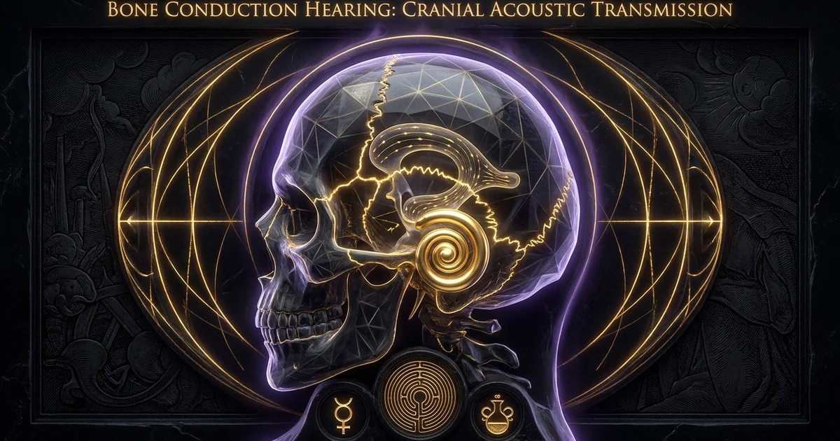
Bone Conduction Hearing: Cranial Acoustic Transmission
Executive Summary & Theoretical Thesis
Non-Ossicular Auditory Transduction Paradigms
Classical psychoacoustics posits the human auditory pathway as an airborne pressure-reception apparatus mediated almost exclusively by the tympano-ossicular chain. In this canonical framework, airborne acoustic waves enter the external auditory meatus, induce mechanical deflection of the tympanic membrane, and are converted into hydraulic displacement within the fluid-filled cochlea via the lever mechanics of the malleus, incus, and stapes.
However, this airborne-centric model overlooks the phylogenetically older and biomechanically sophisticated alternate pathway: bone conduction hearing acoustic transmission through the skull directly to the cochlea. By bypassing the outer and middle ear, mechanical vibrations applied to the cranium propagate through the complex osteological architecture of the head, exciting the basilar membrane and generating sensory transduction identical in neural coding to high-fidelity air conduction.
P_air (Acoustic Field) ---> Tympanum ---> Ossicles ---> Oval Window ---> Cochlear Traveling Wave
                                                                             ^
P_mech (Vibrational Field) ---> Cranial Vault ---> Otic Capsule Matrix -------+
The fundamental assertion of cranial acoustic transmission is that the human neurocranium functions not as a passive, acoustically inert container for encephalic mass, but as an anisotropic, resonant acoustic waveguide. Mechanical perturbations applied to the calvarium induce multi-modal elastodynamic waves that travel across cortical bone plates, traverse complex suture boundaries, and converge upon the petrous portion of the temporal bone.
This non-ossicular pathway operates through an intricate confluence of translational, deformational, and hydroviscous mechanisms, proving that the sensory apparatus of the inner ear maintains an intrinsic, unmediated mechanical impedance link with the global skeletal framework of the human organism.
The Calvarium as an Anisotropic Acoustic Waveguide
The biomechanical efficacy of skull conduction rests upon the structural properties of the calvarium. Far from behaving as an idealized isotropic shell, the human skull represents an inhomogeneous, viscoelastic tri-layer sandwich structure comprising an outer dense cortical plate (lamina externa), a porous central trabecular core filled with diploic marrow (diploë), and an inner dense cortical plate (lamina interna).
The mechanical properties of these layers diverge sharply: the cortical tables exhibit an elastic modulus ($E$) ranging from 10 to 18 GPa and a mass density ($\rho$) approximating $1900\text{ to }2000\text{ kg/m}^3$, whereas the diploë displays an elastic modulus between 0.5 and 3 GPa and a density spanning $1000\text{ to }1500\text{ kg/m}^3$.
[Lamina Externa]  E ≈ 10–18 GPa, ρ ≈ 1900–2000 kg/m³  (Dense Cortical)
---------------------------------------------------------------------
[Diploë Layer]    E ≈ 0.5–3 GPa, ρ ≈ 1000–1500 kg/m³  (Porous Viscoelastic)
---------------------------------------------------------------------
[Lamina Interna]  E ≈ 10–18 GPa, ρ ≈ 1900–2000 kg/m³  (Dense Cortical)
This sandwich architecture establishes a frequency-dependent dispersion profile. At low vibrational frequencies (below approximately 1000 Hz), the cranial vault exhibits standing-wave resonance and rigid-body kinematic dynamics, oscillating along translational and rotational axes with negligible localized phase distortion.
As acoustic excitation exceeds 1 to 1.5 kHz, the wavelength of elastodynamic stress waves compresses below the structural dimensions of the cranial perimeter. Under these short-wavelength regimes, the calvarium transitions into a distributed-parameter waveguide supporting flexural, compressional, shear, and surface wave propagation.
The viscoelastic properties of the diploic marrow induce significant shear deformation and viscoelastic damping, which limits chaotic modal resonances while guiding acoustic energy efficiently toward the rigid skull base and the encased otic capsule.
Acoustic Impedance Discrepancies and Boundary Conditions
The transmission of mechanical vibrational energy across biological tissue interfaces is dictated by the principle of acoustic impedance matching:
$$Z = \rho c$$
where $\rho$ represents the mass density of the medium and $c$ represents the complex acoustic phase velocity. Airborne acoustic transmission confronts an extreme impedance barrier at the air-water boundary: air presents a characteristic acoustic impedance of approximately:
$$Z_{\text{air}} \approx 415\text{ Rayl (Pa}\cdot\text{s/m)}$$
whereas the perilymph and endolymph fluids of the labyrinth approximate the impedance of water or saline:
$$Z_{\text{perilymph}} \approx 1.5 \times 10^6\text{ Rayl (1.5 MRayl)}$$
The middle ear acts as a dynamic mechanical impedance transformer, relying on the areal ratio of the tympanic membrane to the stapes footplate and the ossicular lever ratio to bridge this 3600-fold mismatch.
Air Conduction (AC)
- Input Medium: Airborne longitudinal barometric pressure oscillations ($Z \approx 415\text{ Rayl}$).
- Mechanical Path: External auditory meatus $\to$ tympanic membrane $\to$ malleus-incus-stapes complex $\to$ oval window.
- Primary Impedance Barrier: Air-to-perilymph interface; relies entirely on the middle ear transformer ratio ($\approx 20:1$).
- High-Frequency Attenuation: Governed by middle ear compliance, ossicular joint suspension, and tympanic mass limits.
- Dynamic Threshold: 0 dB HL baseline; highly vulnerable to conductive pathologies (e.g., otosclerosis, tympanic perforation).
Bone Conduction (BC)
- Input Medium: Elastodynamic stress waves within the solid osteological matrix ($Z \approx 6.0\text{–}7.5\text{ MRayl}$).
- Mechanical Path: Calvarium $\to$ petrous temporal bone $\to$ otic capsule $\to$ direct basilar membrane displacement.
- Primary Impedance Barrier: Cutaneous-subcutaneous soft tissue dampening; near-unity matching at osseous boundaries.
- High-Frequency Attenuation: Governed by diploic viscoelastic dissipation and cranial suture acoustic scattering.
- Dynamic Threshold: Bypasses conductive interruptions; baseline efficiency elevated by percutaneous osseointegration.
Cranial acoustic transmission circumvents the middle-ear impedance transformer entirely. Compact cortical bone possesses a characteristic acoustic impedance of roughly:
$$Z_{\text{bone}} \approx 6.0\text{ to }7.5\text{ MRayl}$$
While there is still an acoustic impedance disparity between the petrous temporal bone and the perilymphatic fluid of the scala vestibuli and scala tympani, the transmission coefficient across this solid-fluid interface is orders of magnitude higher than an unassisted air-water boundary.
High-energy mechanical waves applied to the skull exploit this coupling, transferring stress tensors across the dense otic capsule to induce basilar membrane displacement without requiring middle ear integrity.
Historical Lineage & Experimental Precedents
Renaissance Diagnostics: Cardano, Ingrassia, and Capivacci
The formal clinical and physical recognition that the mammalian sensorium can register sound through cranial pathways without the mediation of the tympanum originated during the Renaissance. The earliest explicit documentation appears in the writings of the Italian polymath Girolamo Cardano in his monumental treatise De Subtilitate Libri XXI (1550).
Cardano observed that acoustic information could be transmitted to the sensorium by clutching an iron rod or dagger between the teeth and resting the opposite tip against a vibrating virginal or lute. Cardano correctly deduced that the teeth and mandible serve as solid mechanical conduits that funnel mechanical vibrations into the inner cranial recesses, effectively bypassing any obstruction in the external ear canal.
Vibrating Source (Lute/Virginal)
       │ (Mechanical Contact)
       ▼
Rigid Coupling (Iron Rod / Dagger)
       │ (Axial Elastic Stress)
       ▼
Teeth / Mandible Architecture
       │ (Osteological Conduction)
       ▼
Cranial Base & Petrous Temporal Bone
       │ (Solid-Fluid Wave Coupling)
       ▼
Auditory Sensorium (Cochlear Transduction)
Following Cardano, the Sicilian anatomist Giovanni Filippo Ingrassia—the discoverer of the stapes—demonstrated in post-mortem and clinical settings that a vibrating tuning fork placed upon the teeth produced acoustic sensations even when the tympanic membranes were completely destroyed by suppurative disease.
In the late 16th century, Girolamo Capivacci expanded upon these foundational insights by introducing the first formal diagnostic differential between middle-ear conductive hearing loss and inner-ear sensorineural degeneration.
Capivacci applied a vibrating rod connected to a zither directly to the teeth of deaf patients: if the patient perceived the pitch, the pathology was localized to the tympanic apparatus; if the patient remained insensible to the mechanical vibrations, the lesion was identified within the auditory nerve or the labyrinthine substance itself.
Von Békésy’s Phase-Cancellation Benchmarks
The modern biophysical validation of cranial acoustic transmission culminated in the twentieth-century laboratory investigations of Georg von Békésy. Prior to von Békésy’s work, a persistent controversy divided the physiological acoustics community: did bone-conducted vibrations stimulate the basilar membrane through an entirely distinct biomechanical modality, or did they resolve into the same traveling wave dynamics induced by conventional air-conducted sound?
“When the skull is driven into vibration, the basilar membrane is deflected by the very same hydrodynamic pressure differentials that occur during air conduction. By precisely adjusting the relative amplitude and phase of an airborne tone and a bone-conducted tone presented simultaneously, the sensory output can be driven to zero through complete destructive interference at the basilar membrane.”
— Georg von Békésy, Experiments in Hearing (McGraw-Hill, 1960). Confirming the identity of basilar membrane traveling wave envelopes across disparate input pathways.
Von Békésy resolved this dilemma using phase-cancellation experiments. By presenting an airborne pure tone to the tympanic membrane via an earphone while concurrently driving the forehead or mastoid process with an electromechanical vibrator tuned to the identical frequency, he altered the relative phase and amplitude of the two inputs.
He discovered that at precise phase shifts ($\Delta \phi = \pi$) and calibrated amplitudes, the auditory perception of the sound could be extinguished. This proved that regardless of whether the initial energy vector originates as an atmospheric compressional wave or an osteological elastodynamic wave, both modes converge upon an identical final pathway: a transverse, dispersive traveling wave along the basilar membrane that induces mechanical shearing of the inner hair cell stereocilia against the tectorial membrane.
The Evolution of Osseointegrated Coupling: From Rods to BAHA
For centuries following Cardano, practical exploitation of bone conduction was constrained by the presence of the intervening cutaneous and subcutaneous soft tissues. The dermal layer, adipose tissue, and galeal aponeurosis constitute an inhomogeneous, lossy viscoelastic filter that introduces substantial viscoelastic damping.
Conventional transcutaneous bone-conduction transducers—such as classic audiometric bone oscillators or historical spectacles-mounted hearing aids—must compress the skin against the mastoid with static forces exceeding 2 to 5 Newtons to achieve adequate acoustic coupling.
Even with this static force, the skin attenuates acoustic energy at an average rate of 10 to 15 dB per octave above 1000 Hz, with high-frequency attenuation reaching 25 to 30 dB in the speech-critical 3 to 8 kHz spectrum.
[Transcutaneous Coupling]
Electromechanical Driver ──> Skin / Adipose Layer ──> Cortical Bone
                              (15–30 dB Attenuation,
                               Viscoelastic Damping)

[Percutaneous Osseointegrated Coupling (BAHA)]
Electromechanical Driver ──> Pure Titanium Abutment ──> Cortical Bone Matrix
                              (Direct Structural Bond,
                               Zero Intervening Soft Tissue)
This limitation was resolved in 1977 when Swedish surgeon-researcher Per-Ingvar Brånemark and biomedical engineer Bo Håkansson applied the principle of osseointegration to acoustic transduction.
By implanting a pure titanium fixture directly into the cortical bone of the mastoid process and allowing osteoblasts to form a structural bond with the titanium oxide surface, they created the Bone Anchored Hearing Aid (BAHA).
Percutaneous osseointegrated coupling eliminates the skin barrier entirely, matching the source impedance directly to the cranial acoustic wave impedance of the calvarium. This approach extended the operational frequency bandwidth beyond 8 kHz, reduced total harmonic distortion, and confirmed that direct mechanical drive provides clean, broadband elastodynamic excitation throughout the cranial vault.
Mathematical Formalism & Physical Mechanics
Elastodynamic Wave Equations in Cranial Bone
Acoustic wave propagation within the inhomogeneous, viscoelastic medium of the cranial vault is governed by the Navier-Cauchy equations of motion for a continuous elastic medium, augmented by viscoelastic damping tensors. Let $\mathbf{u}(\mathbf{x}, t)$ represent the dynamic displacement field vector at a given coordinate $\mathbf{x}$ within the cranial bone matrix at time $t$. The generalized wave equation is formulated as:
$$\rho \frac{\partial^2 \mathbf{u}}{\partial t^2} = \nabla \cdot \boldsymbol{\sigma} + \mathbf{F}_{\text{ext}}$$
where $\rho$ is the local cranial bone density, $\boldsymbol{\sigma}$ is the dynamic Cauchy stress tensor, and $\mathbf{F}_{\text{ext}}$ represents external electromechanical driving forces.
Because cranial bone exhibits pronounced viscoelasticity, the constitutive relationship between stress $\boldsymbol{\sigma}$ and the infinitesimal strain tensor $\boldsymbol{\varepsilon} = \frac{1}{2}\left[\nabla \mathbf{u} + (\nabla \mathbf{u})^T\right]$ is defined through the Boltzmann superposition integral or, in the frequency domain ($\omega$), via complex viscoelastic moduli:
$$\boldsymbol{\sigma}(\omega) = \lambda^(\omega) \operatorname{tr}(\boldsymbol{\varepsilon})\mathbf{I} + 2\mu^(\omega)\boldsymbol{\varepsilon}$$
where $\lambda^(\omega) = \lambda_1(\omega) + i\omega\eta_\lambda$ and $\mu^(\omega) = \mu_1(\omega) + i\omega\eta_\mu$ are the frequency-dependent complex Lamé parameters accounting for both dynamic storage elasticity and viscous dissipation.
By applying Helmholtz decomposition to the displacement field $\mathbf{u} = \nabla \Phi + \nabla \times \mathbf{\Psi}$ (where $\Phi$ is the scalar dilatational potential and $\mathbf{\Psi}$ is the divergence-free vector rotational potential), the elastodynamic waves within the compact cortical tables separate into compressional (primary, $P$) and shear (secondary, $S$) wave modes:
$$c_P = \sqrt{\frac{\lambda + 2\mu}{\rho}}, \quad c_S = \sqrt{\frac{\mu}{\rho}}$$
For typical human cortical bone parameters ($\rho \approx 1950\text{ kg/m}^3$, Young’s modulus $E \approx 14\text{ GPa}$, Poisson’s ratio $\nu \approx 0.32$):
- Compressional Wave Velocity: $c_P \approx 3100\text{ to }3400\text{ m/s}$
- Shear Wave Velocity: $c_S \approx 1600\text{ to }1800\text{ m/s}$
Within the bounded geometry of the diploic plate (thickness $h \approx 4\text{ to }8\text{ mm}$), these bulk modes couple at the free boundaries of the lamina externa and lamina interna, resolving into dispersive guided Lamb waves (symmetric $S_0$ and anti-symmetric $A_0$ modes) and surface Rayleigh waves:
$$c_R \approx \frac{0.862 + 1.14\nu}{1 + \nu} c_S \approx 1450\text{ to }1650\text{ m/s}$$
The dispersion relation for the flexural anti-symmetric modes ($A_0$), which dominate skull vibrations between 500 Hz and 4000 Hz, dictates that phase velocity scales with the square root of frequency:
$$c_{\text{flex}} = \left(\frac{D}{\rho h}\right)^{1/4} \sqrt{\omega}$$
where:
$$D = \frac{E h^3}{12(1 - \nu^2)}$$
represents the flexural rigidity of the cranial plate. This dispersion confirms that the skull acts as a dispersive mechanical filter, causing broad-spectrum acoustic impulses to separate into frequency components as they traverse the calvarium toward the petrous bone.
The Five Biophysical Modes of Cochlear Excitation
The transition of cranial elastodynamic stress waves into sensory basilar membrane traveling waves occurs through five primary biophysical mechanisms, as formulated by Tonndorf (1966) and expanded by Stenfelt & Goode (2005):
$$X_{\text{BM}}(\omega) = \sum_{k=1}^5 T_k(\omega) \cdot F_{\text{in}}(\omega)$$
where $X_{\text{BM}}$ is basilar membrane displacement, $F_{\text{in}}$ is the input mechanical driving force, and $T_k(\omega)$ represents the transfer function for each pathway.
                     ┌──> 1. Cochlear Fluid Inertia (dominant < 1 kHz)
                     │
                     ├──> 2. Skull Bone Matrix Distortion (dominant > 1–1.5 kHz)
Cranial Energy Vector ├──> 3. Middle Ear Ossicular Inertial Lag
                     │
                     ├──> 4. Sound Radiation into External Canal (Cartilage)
                     │
                     └──> 5. Intracranial CSF Pressure Coupling (Third Windows)
- Inertia of the Cochlear Fluid: The perilymph mass inside the labyrinth has finite inertia. When translational bone vibrations oscillate the petrous pyramid, the fluid lags behind the surrounding osseous walls. Because the compliant round window membrane possesses much higher acoustic admittance than the stapes footplate seated in the oval window, an asymmetric hydrodynamic pressure gradient develops across the basilar membrane, driving the traveling wave. This mechanism dominates at low frequencies ($< 1000\text{ Hz}$).
- Compressional Distortion of the Otic Capsule: At frequencies exceeding 1 to 1.5 kHz, the cranial acoustic wavelength becomes shorter than the skull’s circumference, producing deformational stress waves. The petrous pyramid experiences cyclic volumetric compression and expansion. Because the scala vestibuli possesses a larger anatomical volume than the scala tympani, equal volumetric compression forces a net hydrodynamic displacement of perilymph from the scala vestibuli toward the scala tympani, directly deflecting the basilar membrane.
- Ossicular Inertial Lag: The ossicles are suspended within the middle ear cavity by ligaments and tendons. When the temporal bone oscillates, the ossicular chain—primarily the mass of the incus and malleus—lags behind the petrous bone motion due to inertia. This relative displacement is transmitted through the stapes footplate into the oval window, generating an unmediated air-conduction-like input.
- Acoustic Radiation into the External Auditory Canal: Elastodynamic waves passing through the temporal bone deform the osseous and cartilaginous walls of the external ear canal. This radiating sound pressure enters the open canal, reflects off the tympanic membrane, and drives the middle ear through conventional air conduction. When the canal is unoccluded, this energy escapes into the environment; when occluded, it redirects into the cochlea, producing the clinical occlusion effect.
- Intracranial Fluid Pressure Transmission: Fluctuations in intracranial pressure induced by cranial vibration propagate through the cerebrospinal fluid (CSF) in the subarachnoid space. These hydroacoustic oscillations enter the labyrinth via the cochlear aqueduct, the endolymphatic duct, and perivascular/perineural channels along the internal auditory canal, acting as a non-osseous hydraulic driver.
Fluid-Structure Boundary Dynamics at the Otic Capsule
The interface between the petrous temporal bone and the labyrinthine perilymph is characterized by fluid-structure interaction (FSI) equations. The acoustic pressure field $p(\mathbf{x}, t)$ within the perilymph satisfies the acoustic wave equation:
$$\nabla^2 p - \frac{1}{c_f^2}\frac{\partial^2 p}{\partial t^2} = 0$$
where $c_f \approx 1500\text{ m/s}$ is the sound speed in perilymph. At the promontory wall and throughout the labyrinthine boundary $\Gamma_{\text{otic}}$, momentum conservation dictates the boundary condition:
$$\mathbf{n} \cdot \nabla p = -\rho_f \mathbf{n} \cdot \frac{\partial^2 \mathbf{u}}{\partial t^2}$$
where $\mathbf{n}$ is the inward normal unit vector to the otic wall, $\rho_f \approx 1000\text{ kg/m}^3$ is the perilymph fluid density, and $\mathbf{u}$ is the elastodynamic displacement vector of the petrous bone.
The promontory acceleration vector, $\mathbf{a}_{\text{prom}} = \frac{\partial^2 \mathbf{u}}{\partial t^2}$, serves as the direct boundary condition driving perilymph displacement. Because the acoustic impedance of the petrous bone ($Z_s \approx 7.0\text{ MRayl}$) and that of the fluid ($Z_f \approx 1.5\text{ MRayl}$) are finite and complex, the pressure transmission across this solid-fluid interface is defined by the normal acoustic transmission coefficient:
$$T_{\text{acoustic}} = \frac{2 Z_f}{Z_f + Z_s}$$
Yielding $T_{\text{acoustic}} \approx 0.35$. Unlike an air-water boundary where transmission is limited to $T \approx 0.001$, more than a third of the boundary normal stress couples into hydraulic pressure, producing basilar membrane deflections across both physiologic and clinical regimes.
Empirical Evidence & Observational Data
3D Laser Doppler Vibrometry of Cranial Modal Resonances
The biomechanical validation of frequency-dependent cranial deformation relies on multi-axis, three-dimensional Laser Doppler Vibrometry (3D-LDV). Contemporary investigations on human cadaveric specimens and in vivo subjects confirm that skull motion undergoes a distinct bifurcation near 1000 Hz.
Frequency Regime 0 Hz ────────── 1000 Hz ────────────────────── 10,000 Hz
Vibrational Mode:  [ Rigid-Body Kinematics ]    [ Structural Modal Resonances ]
Characteristics:   Whole-skull translation/     Complex cymatic nodal lines,
                   rotation; negligible       localized deformation, Lamb/
                   local phase gradient.        Rayleigh wave propagation.
Below 1000 Hz, the cranium vibrates primarily as an intact, rigid mass with six uncoupled degrees of freedom (three translational axes, three rotational axes). Relative deformations between separate cranial bones are minimal, with phase shifts between the driving point and the contralateral promontory remaining below 0.1 radians.
Experimental LDV data demonstrates that between 1.2 kHz and 1.8 kHz, the human skull enters its fundamental structural modal resonances. Point mechanical impedance measurements at the mastoid reveal a characteristic dip in mechanical impedance magnitude $|Z_m|$ from $200\text{ N}\cdot\text{s/m}$ at 200 Hz down to $10\text{–}20\text{ N}\cdot\text{s/m}$ at the first resonance frequency ($\approx 1.4\text{ kHz}$), accompanied by zero-crossings in phase.
— Stenfelt & Goode, Bone-conducted sound: Physiological and clinical aspects, Otol. Neurotol. (2005); Håkansson et al., The mechanical impedance of the human skull, Chest (1986).
Above 1.5 kHz, 3D-LDV mapping reveals complex cymatic modal nodes and nodal antinode lines arrayed across the parietal, occipital, and frontal calvaria.
As the drive frequency approaches 4 to 8 kHz, the distance between nodal lines decreases to centimeters, matching flexural wavelengths ($\lambda_{\text{flex}} \approx 2\text{ to }5\text{ cm}$). Promontory acceleration measurements demonstrate that the otic capsule moves along non-linear elliptical orbits, with the orientation of these orbits shifting as the input excitation changes phase and direction.
Transfer Functions: Percutaneous vs. Transcutaneous BAHA Coupling
Quantifying transmission efficiency requires measuring the promontory acceleration transfer function (PATF), defined as the ratio of promontory acceleration to the dynamic driving force applied at the mastoid:
$$\text{PATF}(\omega) = \frac{|\mathbf{a}{\text{prom}}(\omega)|}{|F{\text{drive}}(\omega)|} \quad \left[\frac{\text{m/s}^2}{\text{N}}\right]$$
PATF [dB rel 1 m/s²/N]
 20 |                                   ... Percutaneous (Direct Bone)
 10 |                 /\  /\           /
  0 |       /\       /  \/  \  /\   ../
-10 |  ____/  \_____/        \/  \./
-20 | /                           \__- - - Transcutaneous (Through Skin)
-30 |/                                   (10–15 dB/octave roll-off)
    +----------------------------------------> Frequency [kHz]
    0.1      0.5     1.0     2.0     5.0    10.0
Comparative biomechanical studies evaluate this transfer function under two conditions: transcutaneous coupling through the intact skin and percutaneous coupling directly to a titanium osseointegrated fixture.
Across the frequency range from 100 Hz to 1000 Hz, both systems exhibit similar baseline acceleration responses, as the skull’s bulk inertial mass dominates overall mechanics. However, above 1 kHz, their performance diverges:
- Percutaneous (Direct Osseointegrated): Maintains an acceleration efficiency between $-5\text{ dB}$ and $+15\text{ dB}$ relative to $1\text{ m/s}^2/\text{N}$ through 10 kHz, capturing the full resonance profile of the temporal bone.
- Transcutaneous (Across Skin): Exhibits a continuous roll-off rate of 10 to 15 dB per octave above 1 kHz. By 4 to 8 kHz, promontory acceleration drops by 20 to 30 dB relative to direct osseointegrated coupling.
This divergence confirms that the soft tissue acts as a low-pass viscoelastic mechanical filter, dissipating high-frequency mechanical energy as heat before it can reach the cortical bone.
Intracranial Hydrodynamics and Dura Mater Coupling
Empirical hydroacoustic measurements in both human cadavers and animal models demonstrate that non-osseous pathways contribute substantially to bone-conduction hearing. When an electromechanical driver stimulates the calvarium, miniature hydrophones situated within the subarachnoid space and lateral ventricles record dynamic intracranial pressure (ICP) swings ranging from 0.1 to 1.5 Pa per Newton of applied driving force.
These oscillatory intracranial pressures travel through non-osseous conduits:
- The Cochlear Aqueduct: Directly bridges the subarachnoid perilymphatic space with the scala tympani, transmitting low-frequency hydroacoustic fluctuations ($< 1\text{ kHz}$) with minimal attenuation.
- The Internal Auditory Canal (IAC): Transmits high-frequency pressure waves along the perineural sheaths of the eighth cranial nerve, communicating through porous cribriform areas into the fundus of the labyrinth.
These findings show that bone conduction does not depend solely on osseous conduction through the bone matrix; it relies equally on hydroacoustic coupling through the encephalic mass, CSF, and dural membranes, which transfer oscillatory pressures into the labyrinth.
Metaphysical Implications & Unified Synthesis
The Cranium as a Sacred Cymatic Cavity
Recognizing the cranium as an anisotropic, viscoelastic waveguide bridges the physical acoustics of bone conduction with historical and esoteric traditions of sacred vocalization. In many classical lineages—such as Vedic recitation, Tibetan tantric chant, and ancient Mediterranean mysteries—vocalization is practiced not merely to project sound into the air, but to generate internal resonance fields within the speaker’s own skull.
When examining internal acoustic stimulation, the cranium can be analyzed as a closed cymatic resonant cavity. As demonstrated in cymatic patterns sound matter geometry, physical substrates subjected to acoustic vibration self-organize along nodal and antinodal boundaries.
The cranium, with its alternating plates of compact bone and diploë, its complex suture lines, and its fluid-filled ventricles, establishes a three-dimensional standing-wave resonator. Sustained internal phonation sets up mechanical wave modes within the sphenoid, ethmoid, and temporal bones, matching the physics of acoustic levitation standing waves within a bounded solid-fluid medium.
Vocal-Somatic Feedback: Mantric Phonation and Sphenoid Tuning
During vocalization, sound pressure within the nasopharynx and laryngeal tract drives the bones of the skull base directly, bypassing the outer ear and the attenuating middle ear acoustic reflex. The stapedius muscle contracts during self-generated phonation to reduce ossicular chain compliance by up to 20 dB, protecting the cochlea from low-frequency saturation.
However, because this reflex relies primarily on middle-ear mechanics, it has little impact on bone-conducted vibrations that bypass the ossicular chain. Self-generated phonation therefore couples directly through the palatine, vomer, and sphenoid bones into the petrous temporal bone:
Vocal Tract Acoustic Resonance
       │
       ▼ (Direct Osseous Contact)
Vomer, Palatine, & Ethmoid Bones
       │
       ▼ (Articular Coupling)
Sphenoid Body & Pterygoid Processes
       │
       ▼ (Basilar Synchondrosis)
Petrous Temporal Bone & Otic Capsule
       │
       ▼ (Direct Hydroacoustic Drive)
Cochlear Fluid & Basilar Membrane
The sphenoid bone occupies a unique architectural position at the center of the skull base, articulating with the frontal, parietal, temporal, occipital, ethmoid, vomer, and zygomatic bones. It forms both the anterior floor of the middle cranial fossa and the sella turcica, which houses the pituitary gland.
Sustained vocal chanting, particularly rich in overtone harmonics (1 to 4 kHz), aligns directly with the natural structural resonance modes of the adult human calvarium. These elastodynamic stress fields oscillate the sphenoid bone, inducing micro-mechanical shear forces against the adjacent dura mater, the cavernous sinus, and the third ventricle, demonstrating how somatic vocal practices produce measurable mechanical effects within deep cranial structures.
Non-Auditory Sensorimotor Integration and Altered Coherence
Beyond its role in basic hearing, the skull serves as a distributed mechanoreceptive array. Cranial acoustic waves stimulate not only the organ of Corti within the cochlea, but also the vestibular system’s otolith organs (the utricle and saccule), which respond directly to mechanical bone vibration.
Acoustic vibrations applied to the cranium between 100 and 1000 Hz evoke Vestibular Evoked Myogenic Potentials (VEMP), showing that cranial acoustic waves stimulate the saccular and utricular maculae directly through bone pathways.
This dual activation of the cochlear and vestibular systems through bone conduction provides a physiological foundation for the shifts in consciousness reported during sustained ritual phonation.
Elastodynamic waves traveling across the cranium stimulate the vestibular nuclei, the reticular activating system, and the vagal autonomic network simultaneously, coupling mechanical pressure variations to neuroelectrical rhythms. Similar vibrational dynamics appear in the design of ancient stone enclosures, as detailed in archaeoacoustic resonance megalithic chambers.
The cranium acts as a physical acoustic transducer, converting skeletal acoustic vibrations into coherent neurological and endocrine responses, and bridging the divide between physical acoustics and somatic physiology.
Frequently Asked Questions
Interaural Attenuation in Cranial Conduction
Question: Why does bone-conducted acoustic energy exhibit negligible interaural attenuation compared to air-conducted sound?
Answer: Interaural attenuation defines the loss of acoustic energy as a signal crosses the head from the ipsilateral test ear to the contralateral non-test ear. For air-conducted sounds delivered via insert earphones or supra-aural headphones, the interaural attenuation ranges from 40 dB to over 70 dB, because the acoustic energy must escape into the atmosphere and wrap around the geometry of the head to reach the opposing ear.
In bone conduction, the cranium behaves as a continuous elastic structure. When a mechanical transducer excites one mastoid, the induced elastodynamic waves propagate across the calvarium with minimal energy dissipation. Consequently, the interaural attenuation for bone-conducted signals ranges from 0 to 15 dB across the conventional audiometric spectrum (250 Hz to 8000 Hz).
Below 1000 Hz, where the skull vibrates as a rigid body, the attenuation frequently approaches 0 dB: both cochleae experience nearly identical mechanical excitation amplitudes.
Above 1500 Hz, wave dispersion, suture damping, and phase interference can produce localized interaural intensity differences of 10 to 15 dB. Because both cochleae are stimulated almost simultaneously, clinical bone-conduction audiometry requires masking the non-test ear with narrowband airborne noise to isolate the ear of interest.
The Third Window Effect and Fluid Inertia
Question: How do anatomical fenestrations or “third windows” within the otic capsule alter the fluid mechanics of bone conduction hearing?
Answer: Normal cochlear reception depends on a dual-window system: the compliant round window balances the motion of the stapes footplate at the oval window, allowing incompressible perilymph to displace back and forth across the basilar membrane.
When a pathological or natural third opening forms—such as in Superior Semicircular Canal Dehiscence (SSCD)—an acoustic “third window” is introduced into the labyrinthine system.
Normal Two-Window System:
[Oval Window]  <====== Perilymph Hydraulic Flow ======> [Round Window]
                               │
                       [Basilar Membrane]
Third Window System (e.g., SSCD):
[Oval Window]  <===+=== Perilymph Flow ===+===> [Round Window]
│                      │
▼                      │
[Dehiscence / "Third Window"]    ▼
(Hydrodynamic Shunt into CSF)   [Basilar Membrane]
This third window alters the hydrodynamics of air and bone conduction in opposite directions:
- For Air Conduction: The dehiscence serves as an acoustic shunt, allowing airborne acoustic energy entering the oval window to escape into the intracranial cavity rather than deflecting the basilar membrane, which elevates air-conduction thresholds (conductive hearing loss).
- For Bone Conduction: The dehiscence lowers the acoustic impedance on one side of the labyrinth, increasing the impedance mismatch between the two sides of the basilar membrane. When the skull vibrates, this increased asymmetry amplifies the net hydrodynamic flow of perilymph across the basilar membrane.
Consequently, patients with SSCD exhibit bone-conduction thresholds that surpass normal basilar membrane sensitivity (dropping below 0 dB HL), frequently reporting audible sensations from intrinsic physiological sounds, including ocular movements, heartbeat, and musculoskeletal motion.
Suture Line Scattering and Wave Dispersion
Question: What role do cranial sutures play in scattering and dampening high-frequency elastodynamic stress waves?
Answer: Cranial sutures—including the coronal, sagittal, lambdoid, and squamosal sutures—are unossified syndesmoses composed of collagenous Sharpey’s fibers and connective tissue matrices that join adjacent osteological plates. From an acoustic perspective, sutures function as structural impedance discontinuities:
$$Z_{\text{suture}} \ll Z_{\text{cortical}}$$
When high-frequency flexural and shear waves encounter these interfaces, the lower elastic modulus of the fibrous suture scatters the wave energy. This scattering induces:
- Wave reflection back into the source cranial plate.
- Mode conversion (e.g., compressional-to-shear transition).
- Viscoelastic dissipation of wave amplitude into heat.
At low frequencies ($< 1000\text{ Hz}$), the dynamic wavelength is much larger than the suture’s structural width, allowing stress waves to pass across the boundary with minimal scattering.
However, at frequencies above 2 to 3 kHz, where flexural wavelengths shorten toward the scale of individual bones, the sutures act as low-pass mechanical filters. They isolate regional skull vibrations, prevent sharp resonance peaks, and damp the overall cranial cavity into a controlled, stable resonator. This complex dispersion dynamic relates closely to the wave mechanics detailed in scalar wave mechanics. :::