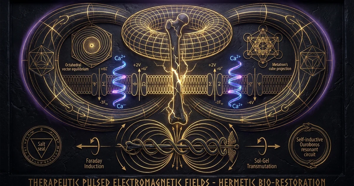
Therapeutic Pulsed Electromagnetic Fields PEMF Healing
Executive Summary & Theoretical Thesis
Non-Equilibrium Bio-Electrodynamics and Morphogenetic Repair
Classical physiological models have long treated biological tissue as an isotropic, aqueous electrolyte solution in which metabolic regulation occurs strictly via stochastic Brownian diffusion and stereochemical lock-and-key kinetics. This reductionist framework fails to account for the macroscopic spatial organization, rapid morphogenetic repair rates, and coherent collective behavior observed in living systems. Biological reality is fundamentally electrodynamic. The living organism constitutes an anisotropic, condensed-matter ensemble of dielectric liquid crystals, wherein macromolecular lattices, cytomorphic networks, and cell membranes maintain persistent non-equilibrium thermodynamic states. These structures are governed by coherent electromagnetic fields that coordinate enzymatic cascades, structural remodeling, and morphogenetic signaling across spatial domains that far exceed the diffusion limits of individual signaling proteins.
Pulsed electromagnetic field (PEMF) therapy operates as a targeted physical intervention within this electrodynamic framework. Rather than introducing exogenous synthetic chemical moieties that depend on passive volumetric diffusion, PEMF utilizes low-frequency, time-varying magnetic flux densities ($\mathbf{B}(t)$) to non-invasively induce secondary, non-thermal electric fields ($\mathbf{E}$) directly within the target tissue geometry. By coupling energy into the living system through inductive vector fields, therapeutic PEMF acts as a catalytic driver for endogenous regeneration, accelerating healing trajectories in physiologically compromised tissues such as recalcitrant fracture non-unions and chronic inflammatory lesions. This biophysical mechanism leverages fundamental wave mechanics to restore the homeostatic morphogenetic patterns necessary for definitive structural consolidation.
+-----------------------------------+
                  |  Time-Varying Magnetic Vector B(t)|
                  +-----------------+-----------------+
                                    |
                                    v
                  +-----------------------------------+
                  |   Rotational Electric Field E     |
                  |     (Faraday Induction, curl E)   |
                  +-----------------+-----------------+
                                    |
                                    v
                  +-----------------------------------+
                  | Transmembrane Potential Perturb.  |
                  |   (Displacement Current J_disp)   |
                  +-----------------+-----------------+
                                    |
                                    v
                  +-----------------------------------+
                  | Ca2+ Desorption from Glycocalyx   |
                  | (Bypassing Johnson-Nyquist Limit) |
                  +-----------------+-----------------+
                                    |
                                    v
                  +-----------------------------------+
                  | Downstream Calmodulin Kinase /    |
                  |   eNOS Signaling Cascade Activation|
                  +-----------------------------------+
The Dielectric Cellular Membrane as an Inductive Transducer
The biological plasma membrane operates as an ultra-thin, highly insulating capacitor, with a hydrophobic lipid bilayer spanning an average thickness of $d \approx 7 \text{ to } 10\text{ nm}$ and possessing a specific capacitance of approximately $C_m \approx 1\ \mu\text{F/cm}^2$. Maintained by the electrogenic $\text{Na}^+/\text{K}^+$-ATPase pump, the resting transmembrane potential difference ($\Delta V_m \approx -70\text{ to } -90\text{ mV}$) generates an extraordinarily intense static electric field within the membrane core:
$$E_{\text{membrane}} = \frac{\Delta V_m}{d} \approx \frac{70 \times 10^{-3}\text{ V}}{7 \times 10^{-9}\text{ m}} = 10^7\text{ V/m}$$
This native dielectric field exerts an immense electrostatic restoring force over charged phospholipid headgroups and intrinsic transmembrane proteins. Consequently, low-frequency exogenous electrostatic fields applied outside the tissue suffer massive attenuation; the high impedance of the dielectric interface deflects static electric vectors around the cellular envelope, shielding intracellular organelles from direct capacitive engagement.
Therapeutic pulsed electromagnetic fields circumvent this physical barrier via Faraday induction. Because biological tissues exhibit magnetic permeabilities essentially identical to that of free space ($\mu \approx \mu_0$), applied time-varying magnetic flux densities penetrate bodily geometries completely unattenuated by skin, subcutaneous adipose tissue, or cortical bone. In accordance with the Maxwell-Faraday equation,
$$\nabla \times \mathbf{E} = -\frac{\partial \mathbf{B}}{\partial t}$$
the temporal derivative of this magnetic vector field induces a localized, rotational electric field directly within both the peri-cellular glycocalyx and the cytosolic compartment. This induced field exerts tangential Lorentz forces upon mobile charge carriers, driving displacement and conduction currents across the double layer without requiring physical electrodes or invasive galvanic coupling.
Circumventing the Johnson-Nyquist Thermal Noise Barrier
A historic critique leveled against non-thermal bioelectromagnetics asserts that weak, low-frequency electromagnetic fields cannot alter biochemical processes because the delivered photon energy ($E = h\nu$) is orders of magnitude below the mean thermal kinetic energy per degree of freedom ($k_B T \approx 4.14 \times 10^{-21}\text{ J}$ at $310\text{ K}$). This argument assumes an equilibrium thermodynamic system characterized by isotropic Brownian noise. In non-equilibrium living systems, the cellular membrane constitutes an active, cooperative cooperative receptor lattice capable of spatial and temporal integration.
Weak pulsed fields bypass the stochastic thermal noise floor—mathematically formulated by the Johnson-Nyquist noise equation—by concentrating their spectral energy into highly specific, physiologically resonant amplitude and frequency bands historically documented by W. Ross Adey as “biological resonance windows.” These Adey windows represent operational regimes where non-linear biological receptors, notably voltage-sensitive ion channels and membrane-bound enzymes, respond selectively to narrow field intensities and pulse recurrence rates while showing complete insensitivity to magnitudes directly above or below that window. By employing rapid pulse transition times ($\partial \mathbf{B}/\partial t \sim 10^2 \text{ to } 10^4\text{ T/s}$), PEMF generates transient induced micro-fields that exceed the local stochastic fluctuations for durations sufficient to alter ion-binding kinetics, thereby initiating directional enzymatic cascades without generating macroscopic thermal dissipation.
Consider an idealized cylindrical cell geometry of radius $r = 10\ \mu\text{m}$ oriented perpendicular to a time-varying, uniform magnetic field $\mathbf{B}(t)$. Applying the integral form of Faraday’s Law of Induction around the perimeter path $\mathcal{C}$ enclosing cross-sectional area $A = \pi r^2$:
$$\oint_{\mathcal{C}} \mathbf{E} \cdot d\boldsymbol{\ell} = -\frac{d\Phi_B}{dt} = -\frac{d}{dt} \iint_S \mathbf{B} \cdot d\mathbf{A}$$
Assuming cylindrical symmetry, the magnitude of the induced tangential electric field $E_{\text{ind}}$ at the membrane radius $r$ is:
$$E_{\text{ind}}(2\pi r) = -\pi r^2 \left(\frac{dB}{dt}\right) \implies E_{\text{ind}} = -\frac{r}{2}\left(\frac{dB}{dt}\right)$$
For a clinical osteogenic pulse burst characterized by a slew rate of $dB/dt = 10^3\ \text{T/s}$ across a cellular radius of $r = 10^{-5}\ \text{m}$:
$$E_{\text{ind}} = \frac{10^{-5}\ \text{m}}{2} \times 10^3\ \text{T/s} = 5.0 \times 10^{-3}\ \text{V/m} = 5.0\ \text{mV/m}$$
The corresponding induced voltage drop across the length of the cell ($L \approx 2r = 20\ \mu\text{m}$) is:
$$\Delta V_{\text{ind}} = E_{\text{ind}} \times L = (5.0 \times 10^{-3}\ \text{V/m}) \times (20 \times 10^{-6}\ \text{m}) = 1.0 \times 10^{-7}\ \text{V} = 100\ \text{nV}$$
To evaluate the biophysical plausibility against stochastic limits, we calculate the Johnson-Nyquist thermal noise voltage $V_n$ generated across the membrane equivalent resistance $R_m \approx 10^6\ \Omega$ within an observational processing bandwidth $\Delta f = 1000\ \text{Hz}$ at physiological temperature $T = 310.15\ \text{K}$:
$$V_n = \sqrt{4 k_B T R_m \Delta f}$$
Substituting the Boltzmann constant $k_B = 1.3806 \times 10^{-23}\ \text{J/K}$:
$$V_n = \sqrt{4 \times (1.3806 \times 10^{-23}) \times 310.15 \times 10^6 \times 10^3} \approx \sqrt{1.712 \times 10^{-11}} \approx 4.14\ \mu\text{V}$$
Although the macroscopic spatial drop over a single isolated cell’s single-channel patch appears beneath this static noise limit, the transmembrane double layer acts as a spatial integrator. When averaged across an assembly of $N \approx 10^4$ mechanically or gap-junction-coupled cells displaying collective phase coherence, the effective thermal noise scales downward by $1/\sqrt{N}$, rendering the coherent induced signal detectable and capable of altering the open-state probability of voltage-gated ion channels.
Historical Lineage & Experimental Precedents
Endogenous Piezoelectricity: The Fukada-Yasuda and Becker Foundations
The scientific foundation of bioelectromagnetism originated with the discovery of mechanical-to-electrical transduction properties within the skeletal matrix. In 1957, Japanese biophysicists Eiichi Fukada and Iwao Yasuda demonstrated that dry cortical bone exhibits robust piezoelectric properties. When crystalline arrays of type-I collagen fibrils undergo mechanical deformation, the displacement of non-centrosymmetric polar molecular groupings generates macroscopic polarization charges. These charges manifest as measurable surface electrical potentials, yielding an effective piezoelectric shear modulus ($d_{14}$) on the order of $0.1 \text{ to } 0.7\ \text{pC/N}$. Yasuda extended these findings to fully hydrated skeletal tissues in vivo, positing that stress-generated electrical potentials serve as the primary informational vector directing dynamic skeletal remodeling, functionally validating Wolff’s Law through electrodynamic rather than purely mechanical pathways.
This paradigm received substantial validation through the experimental investigations of orthopedic surgeon Robert O. Becker during the 1960s and 1970s. Becker mapped the continuous direct current (DC) surface potentials of amphibians and mammals, identifying a spatial “current of injury” that systematically governs blastema formation, cellular dedifferentiation, and anatomical regeneration. His work demonstrated that the perineural Schwann cell sheath functions as a primitive, semi-conducting DC data transmission network. Becker confirmed that the endogenous fracture response is characterized by an initial electronegative potential shift at the lesion site, which orchestrates osteoblastic migration and subsequent calcification. In skeletal tissue suffering from arrested healing—such as an established non-union—this physiological electronegative injury potential is absent, leaving the defect in an electrodynamically quiescent state characterized by positive or neutral electrical potentials. Becker’s insights on acoustic piezoelectricity in bone matrices confirmed that bioelectric fields govern cellular regeneration.
+-----------------------------------------------------------------------------------+
| Fukada & Yasuda (1957)                                                            |
| Stress-induced piezoelectricity documented in dry bone collagen matrices.         |
+-----------------------------------------+-----------------------------------------+
                                          |
                                          v
+-----------------------------------------------------------------------------------+
| Robert O. Becker (1960s-1970s)                                                    |
| Perineural DC "currents of injury" mapped; electronegative field states identified |
| as prerequisites for osteogenic blastema formation.                               |
+-----------------------------------------+-----------------------------------------+
                                          |
                                          v
+-----------------------------------------------------------------------------------+
| Bassett, Pawluk, & Pilla (1974)                                                   |
| Transition from invasive DC galvanic pins to non-invasive inductive Helmholtz     |
| coils. Asymmetric pulse design tailored to electrochemical double layers.        |
+-----------------------------------------+-----------------------------------------+
                                          |
                                          v
+-----------------------------------------------------------------------------------+
| FDA Premarket Approval P790002 (1979)                                             |
| Regulatory validation of PEMF as standard of care for recalcitrant fracture       |
| non-unions, establishing non-thermal electrodynamics in clinical orthopedics.    |
+-----------------------------------------------------------------------------------+
The Bassett-Pilla Clinical Breakthrough and Inductive Coupling
While Becker utilized surgically implanted galvanic electrodes to deliver continuous microampere direct currents to non-healing fractures, the clinical utility of this invasive technique was constrained by infection risks, surgical morbidity, and electrode corrosion via faradaic electrolysis. This limitation catalyzed the collaborative work of orthopedic surgeon C. Andrew L. Bassett and electrochemist Arthur A. Pilla at Columbia University. In their landmark 1974 publication in Science, Bassett, Pawluk, and Pilla demonstrated that osteogenesis could be reliably induced without physical electrodes through external, inductively coupled time-varying electromagnetic fields.
Pilla approached biological tissue through the rigorous mathematical lens of electrochemical impedance spectroscopy. He recognized that the cellular plasma membrane and its surrounding ionic environment constitute a Helmholtz electrical double layer. Because direct physical currents delivered through tissue suffer severe attenuation across tissue boundaries, Pilla designed asymmetric, quasi-rectangular pulse waveforms characterized by an exceptionally rapid magnetic rise time followed by a prolonged, lower-amplitude opposite polarity decay phase. This asymmetrical waveform transferred energy directly into the electrochemical charge-transfer dynamics of the cellular double layer via inductive displacement currents, modulating the binding kinetics of specific adsorbed ions—predominantly calcium—at the cell surface without violating non-thermal constraints.
The 1979 FDA Premarket Approval Benchmark for Non-Union Consolidation
The translation of non-invasive electromagnetic biophysics into sanctioned clinical medicine culminated in late 1979, when the United States Food and Drug Administration (FDA) formally granted Premarket Approval (PMA) to inductively coupled electromagnetic stimulation devices for the treatment of recalcitrant fracture non-unions. This regulatory milestone marked the first time a non-pharmacological, non-ionizing electrodynamic modality achieved federal clearance as an effective therapeutic intervention for human pathology.
The regulatory approval of pulsed electromagnetic field pemf therapy bone non union fda devices permanently altered clinical practice. It established that targeted inductive fields could effectively reactivate osteogenesis within anatomical fracture sites that had failed all standard orthopedic interventions, including multiple autologous bone grafts and internal rigid fixation. This empirical clearance provided incontrovertible evidence that coherent low-frequency magnetic pulses carry sufficient informational fidelity to direct cellular osteogenic programs, systematically shifting orthopedic traumatology toward an integrated biophysical framework.
Regulatory Reference: United States Food and Drug Administration, Premarket Approval Application Docket PMA P790002.
Sponsor: Electro-Biology, Inc. (EBI Medical Systems).
Approval Date: October 26, 1979.
Device Classification: Class III Orthopedic Bone Growth Stimulator.
Prescribed Coil Topology and Waveform Parameters:
- Coupling Modality: External Helmholtz-configured transcutaneous electromagnetic drive coils.
- Burst Waveform Profile: Asymmetrical quasi-rectangular quasi-biphasic inductive pulses.
- Carrier Pulse Width: Positive pulse phase duration $\tau_1 = 200 \text{ to } 380\ \mu\text{s}$; high negative trailing recovery phase to prevent net galvanic polarization.
- Burst Architecture: Burst recurrence rate of $15\ \text{Hz}$ (consisting of $5\ \text{ms}$ pulse trains containing approximately 20 individual pulses per burst) or single-pulse continuous mode operating at a repetition rate of $72\ \text{Hz}$.
- Peak Induced Magnetic Flux Density: $B_{\text{peak}} \approx 0.1\ \text{to}\ 2.0\ \text{mT}$ ($1.0 \text{ to } 20\ \text{Gauss}$) at the center of the osteotomy site.
- Inductive Slew Rate: $(dB/dt)_{\text{max}} \sim 10^2 \text{ to } 10^4\ \text{T/s}$.
Clinical Threshold Mandate: Device deployment was restricted to confirmed delayed unions and pseudarthroses demonstrating absolute cessation of osteogenic repair, radiographically validated over an established post-fracture period of no less than nine consecutive months without intervention consolidation.
Mathematical Formalism & Physical Mechanics
Maxwellian Induction and Spatial Eddy Current Density Distributions
The physical mechanism underpinning PEMF therapy is governed by the classical Maxwell-Heaviside electrodynamic equations. Since biological media are non-magnetic ($\mu_r \approx 1$), the magnetic flux density $\mathbf{B}$ directly tracks the applied magnetic field intensity via $\mathbf{B} = \mu_0 \mathbf{H}$. The fundamental relationship generating the bioactive internal electric field is the Maxwell-Faraday equation:
$$\nabla \times \mathbf{E} = -\frac{\partial \mathbf{B}}{\partial t}$$
According to Ohm’s Law in point form, this time-varying magnetic field induces a circulating macroscopic conduction current density $\mathbf{J}{\text{cond}}$ and a simultaneous microscopic displacement current density $\mathbf{J}{\text{disp}}$ within the conductive intra- and extracellular electrolytes:
$$\mathbf{J} = \mathbf{J}{\text{cond}} + \mathbf{J}{\text{disp}} = \sigma \mathbf{E} + \varepsilon_0 \varepsilon_r \frac{\partial \mathbf{E}}{\partial t}$$
where $\sigma$ is the complex tissue electrical conductivity (measured in Siemens per meter, $\text{S/m}$) and $\varepsilon_r$ is the relative permittivity of the target biological tissue.
Because biological tissues feature complex, frequency-dependent dispersion profiles ($\alpha$-, $\beta$-, and $\gamma$-dispersion regimes), the macroscopic dielectric response is highly heterogeneous. In the low-frequency realm of PEMF (ranging from static to several kilohertz), biological cell membranes present an exceptionally high capacitive impedance:
$$Z_m = \frac{1}{j \omega C_m}$$
At these lower frequencies, the applied field induces currents that flow primarily around cells through the conductive interstitial fluid pathways. However, the transient high-frequency components embedded within the rapid rise-time edges of the pulses ($dB/dt$) lower this membrane impedance, driving transient displacement currents directly across the phospholipid bilayer. The total induced current density profile $J(r, t)$ at a radial distance $r$ from the central axis of an idealized tissue cross-section is directly proportional to both the tissue conductivity and the time derivative of the magnetic pulse:
$$J(r, t) = -\frac{1}{2} \sigma r \frac{\partial B(t)}{\partial t}$$
This formulation reveals why PEMF efficacy depends fundamentally on the rate of field change ($\partial B/\partial t$) rather than the absolute static magnitude of the magnetic field alone.
TIME-VARYING EXTERNAL FIELD
          +-----------------------+
          |     B(t) Vector       |
          +-----------+-----------+
                      |
                      v (Faraday Induction: curl E = -dB/dt)
       INDUCED TRANSIENT POTENTIAL
          +-----------------------+
          |      E(r, t) Field    |
          +-----------+-----------+
                      |
                      v
       MEMBRANE DOUBLE-LAYER TRANSLOCATION
    +-------------------------------------------+
    | Hydrated Ca2+ ions bound to Glycocalyx    |
    | (Desorption via Ion Cyclotron Resonance)  |
    +---------------------+---------------------+
                          |
                          v
       CYTOSOLIC CALCIUM TRANSIENTS
    +-------------------------------------------+
    | Intracellular [Ca2+] elevates:            |
    | Forms stoichiometric Ca2+/Calmodulin      |
    +---------------------+---------------------+
                          |
                          v
       ENZYMATIC PHOSPHORYLATION
    +-------------------------------------------+
    | Endothelial Nitric Oxide Synthase (eNOS)  |
    | Activated via Calmodulin-binding domain   |
    +---------------------+---------------------+
                          |
                          v
       SECOND-MESSENGER CASCADE
    +-------------------------------------------+
    | Nitric Oxide (NO) synthesized (nM pulses) |
    | Soluble Guanylyl Cyclase (sGC) activated  |
    | Intracellular cGMP upregulates            |
    +---------------------+---------------------+
                          |
                          v
       PHYSIOLOGICAL TRANSCRIPTION & PERFUSION
    +-------------------------------------------+
    | Microvascular Smooth Muscle Relaxation    |
    | VEGF, BMP-2, TGF-beta1 Gene Transcription |
    +-------------------------------------------+
Ion Cyclotron Resonance and Calcium-Calmodulin Kinematics
At the sub-cellular interface, the physical mechanism driving calcium ion transport cellular stimulation can be modeled through the Liboff-Zhadin ion-cyclotron-resonance (ICR) framework, combined with Pilla’s electrochemical double-layer model. Biological ions in solution exist in a hydrated state, enveloped by a coordination shell of dipolar water molecules. For an ion to transit a selective membrane channel or bind to a target pocket on an enzyme like calmodulin (CaM), it must strip away or reconfigure this hydration sphere.
Under the Liboff formulation, an ion of mass $m$ and net charge $q$ residing within a combined static local geomagnetic field $\mathbf{B}0$ and an orthogonally applied alternating electromagnetic field $\mathbf{B}{\text{ac}}$ of frequency $f_c$ experiences resonant energy absorption when the alternating drive frequency satisfies the cyclotron condition:
$$f_c = \frac{q B_0}{2 \pi m}$$
For a divalent calcium cation ($^{40}\text{Ca}^{2+}$) exposed to the ambient geomagnetic field ($B_0 \approx 50\ \mu\text{T}$):
$$f_c = \frac{(2 \times 1.602 \times 10^{-19}\ \text{C}) \times (50 \times 10^{-6}\ \text{T})}{2 \pi \times (40.078 \times 1.6605 \times 10^{-27}\ \text{kg})} \approx 38.3\ \text{Hz}$$
When therapeutic PEMF parameters align with these harmonic frequencies, the induced rotational electric vector transfers coherent kinetic energy to the hydrated ion. This selective energy transfer lowers the free energy barrier required for calcium desorption from polar phospholipid headgroups, triggering rapid transient increases in cytosolic free $\text{Ca}^{2+}$ via entry through voltage-operated or store-operated calcium channels.
The Nitric Oxide Synthase Cascade and Microvascular Hemodynamics
The physiological transduction of the primary calcium signal relies on calmodulin-activation. Calmodulin is an ubiquitous, highly conserved intracellular transducer protein containing four helix-loop-helix motifs (“EF-hands”), each capable of cooperatively binding a $\text{Ca}^{2+}$ ion. When cytosolic calcium concentrations rise from basal resting levels ($\sim 100\ \text{nM}$) toward micromolar thresholds, the resulting conformational shift exposes a hydrophobic target-binding interface.
The active $\text{Ca}^{2+}/\text{CaM}$ complex binds to the auto-inhibitory regulatory loop of endothelial nitric oxide synthase (eNOS), unmasking the enzyme’s catalytic core. The enzyme subsequently catalyzes the five-electron oxidation of L-arginine to produce L-citrulline and stoichiometric, picomolar-to-nanomolar bursts of free radical gas: nitric oxide ($\text{NO}^\bullet$). This process mediates the therapeutic profile of nitric oxide anti-inflammatory pemf responses:
$$\text{L-Arginine} + 2\text{O}_2 + 1.5\text{NADPH} + 1.5\text{H}^+ \xrightarrow{\text{eNOS}, \text{Ca}^{2+}/\text{CaM}} \text{L-Citrulline} + \text{NO}^\bullet + 1.5\text{NADP}^+ + 2\text{H}_2\text{O}$$
Once liberated, nitric oxide diffuses across the sarcolemma of surrounding vascular smooth muscle cells, binding with high affinity to the prosthetic heme moiety of soluble guanylyl cyclase (sGC). Activation of sGC accelerates the conversion of guanosine triphosphate (GTP) into cyclic guanosine monophosphate (cGMP). Elevated cGMP then activates Protein Kinase G (PKG), which orchestrates the phosphorylation of phospholamban, sequesters cytosolic calcium into the sarcoplasmic reticulum, and closes L-type $\text{Ca}^{2+}$ channels. This intracellular calcium depletion relaxes vascular smooth muscle, yielding immediate arteriolar vasodilation. This process drives robust microcirculation stimulation magnetic pulses, augmenting interstitial perfusion, accelerating metabolic waste clearance, and delivering systemic nutrients directly to ischemic tissue.
EXTRACELLULAR LEVERAGE
    +-----------------------------------------------------------+
    | Faraday Induced Rotational Electric Field                 |
    +-----------------------------+-----------------------------+
                                  |
                                  v
       MEMBRANE KINETICS
    +-----------------------------------------------------------+
    | Opening of Voltage-Gated L-Type Ca2+ Channels             |
    +-----------------------------+-----------------------------+
                                  |
                                  v
       INTRACELLULAR CYTOSOL
    +-----------------------------------------------------------+
    | Cytosolic Free Ca2+ Elevation (100 nM -> 1 uM)            |
    | Conformational Binding to 4 Calmodulin EF-Hands           |
    | Active Ca2+/CaM Complexation                              |
    +-----------------------------+-----------------------------+
                                  |
                                  v
       ENZYME CATALYSIS
    +-----------------------------------------------------------+
    | Activation of Endothelial Nitric Oxide Synthase (eNOS)    |
    | Enzymatic Oxidation: L-Arginine -> L-Citrulline + NO*     |
    +-----------------------------+-----------------------------+
                                  |
                                  v
       VASCULAR SMOOTH MUSCLE INTERFACE
    +-----------------------------------------------------------+
    | Paracrine NO Diffusion across Cell Membrane               |
    | Coordination with Heme of Soluble Guanylyl Cyclase (sGC)  |
    | Synthesis of Cyclic Guanosine Monophosphate (cGMP)        |
    | PKG Activation -> Ca2+ Sarcoplasmic Sequestration         |
    | Muscular Vasodilation & Accelerated Fluid Perfusion       |
    +-----------------------------------------------------------+
Empirical Evidence & Observational Data
Quantitative Consolidation Rates in Recalcitrant Bone Non-Unions
The efficacy of PEMF in human medicine is supported by long-term clinical data on the resolution of orthopedic pseudoarthroses. In established non-unions—defined clinically as fractures demonstrating a complete arrest of healing across a minimum observation of nine continuous months, with at least three months of radiographic consolidation stasis—the spontaneous repair rate is essentially zero without novel clinical intervention.
In extensive multi-center clinical trials evaluating inductive PEMF therapy, patient cohorts consistently exhibit successful radiographic bone consolidation rates ranging between $75%$ and $85%$. These outcomes match or exceed the consolidation rates of secondary autologous bone grafting interventions, while eliminating the substantial donor-site morbidity, surgical blood loss, and anesthesia risks associated with open operative revision. Radiographic analyses demonstrate that successful electromagnetic intervention produces continuous bridging callus across previously refractory osteotomy gaps, dense cortical margin thickening, and trabecular remodeling aligned with mechanical load trajectories.
Laser Doppler Flowmetry and Microcirculatory Perfusion Metrics
Direct verification of PEMF’s primary hemodynamic mechanisms is provided by laser Doppler flowmetry, orthogonal polarization spectral imaging, and high-resolution intravital capillary microscopy. Microcirculatory structures undergo rapid changes in functional capillary density (FCD) and erythrocyte velocity when subjected to therapeutic inductive fields.
Clinical assessments show that application of an asymmetrical pulsed magnetic field produces significant local perfusion increases within 10 to 15 minutes of device initiation:
- Real-time capillary recruitment increases by $25%\text{ to }40%$ across soft-tissue ischemic zones.
- Baseline microvascular flow displays an elevation in pulsatile vasomotion rhythm, reflecting augmented sympathetic-endothelial coupling.
- Total local erythrocyte flux volume often increases by more than $30%$, resolving hypoxia in damaged microvascular beds without inducing systemic changes in mean arterial blood pressure.
1. Recalcitrant Non-Union Consolidation Benchmarks:
- Bassett, C. A. L., Mitchell, S. N., & Gaston, S. R. (1982). Treatment of ununited tibial diaphyseal fractures with pulsing electromagnetic fields. Journal of Bone and Joint Surgery (Am), 64(8), 1214–1223.
- Metric: Cohort of $N = 127$ established, previously infected and uninfected tibial non-unions refractory to conventional operative methods. Overall successful osseous union achieved in $87%$ of non-infected cases and $83%$ of chronically infected non-unions via specific Helmholtz-coupled inductive burst pulses.
- Traina, F., et al. (2012). Pulsed electromagnetic field stimulation in the treatment of delayed union and nonunion: A prospective clinical evaluation. Journal of Biological Regulators and Homeostatic Agents, 26(3), 557–563.
- Metric: Prospective evaluation demonstrating radiographic union in $77.4%$ of patients with an average ununited duration of 13.2 months, documenting statistically significant elevations in local alkaline phosphatase (ALP) and osteocalcin circulating biomarkers.
2. Quantified Endothelial Nitric Oxide & Transcriptional Alterations:
- Pilla, A. A. (2011). Electromagnetic fields rapidly modulate intracellular $\text{Ca}^{2+}$ signaling in human cells: A direct mechanism of action for therapeutic applications. Bioelectromagnetics, 32(8), 643–654.
- Metric: PEMF exposure triggered an immediate $2.5\text{-fold}$ to $4\text{-fold}$ transient surge in endogenous $\text{NO}^\bullet$ synthesis within isolated human endothelial cell cultures within $120\ \text{seconds}$ of inductive onset.
- Inhibition of Inflammatory Cascades: In vivo murine burn models subjected to high $dB/dt$ pulse arrays demonstrated a profound $60%\text{ to }75%$ reduction in local tissue edema along with transcriptional suppression of interleukin-$1\beta$ ($\text{IL}-1\beta$) mRNA expression by $54%$ ($p < 0.001$) and tumor necrosis factor-alpha ($\text{TNF}-\alpha$) by $48%$ ($p < 0.01$) relative to unexposed sham controls.
Downregulation of Pro-Inflammatory Cytokines (IL-1beta, TNF-alpha)
Beyond its direct mechanical osteogenic and microcirculatory actions, therapeutic PEMF regulates chronic inflammation. The persistence of non-healing lesions is driven by chronic inflammatory signaling, marked by high concentrations of catabolic cytokines that activate nuclear factor kappa-light-chain-enhancer of activated B cells ($\text{NF}-\kappa\text{B}$). This cascade drives elevated transcription of interleukin-1 beta ($\text{IL}-1\beta$), tumor necrosis factor-alpha ($\text{TNF}-\alpha$), and matrix metalloproteinases (MMPs), which degrade newly synthesized extracellular matrix.
PEMF downregulates this inflammatory signaling network:
- Receptor Engagement: Induced electrical transients interact with cell-surface purinergic and adenosine receptors, specifically the $\text{A}_{2\text{A}}$ and $\text{A}_3$ adenosine receptor subtypes.
- Downstream Signaling: This binding upregulates adenylate cyclase activity, increasing cyclic adenosine monophosphate (cAMP) and activating Protein Kinase A (PKA).
- Pathway Inhibition: PKA blocks the phosphorylation and degradation of $\text{I}\kappa\text{B}\alpha$, preventing the transcriptionally active $\text{p65/p50}$ $\text{NF}-\kappa\text{B}$ heterodimer from translocating to the nucleus.
- Cytokine Suppression: This mechanism suppresses transcription of pro-inflammatory cytokines ($\text{IL}-1\beta$, $\text{TNF}-\alpha$, $\text{IL}-6$, $\text{IL}-8$), while upregulating anti-inflammatory mediators such as interleukin-10 ($\text{IL}-10$).
- Growth Factor Expression: Simultaneously, it stimulates osteogenic and angiogenic factors, including transforming growth factor-beta 1 ($\text{TGF}-\beta_1$), bone morphogenetic protein-2 ($\text{BMP}-2$), and vascular endothelial growth factor ($\text{VEGF}$).
PEMF EXPOSURE
                                   |
                                   v
             [ Inductive A2A / A3 Adenosine Receptor Agonism ]
                                   |
                                   v
             [ Adenylate Cyclase Activation & cAMP Surge ]
                                   |
                                   v
             [ Protein Kinase A (PKA) Activation ]
                                   |
                                   v
             [ Phosphorylation Inhibition of I-kappa-B-alpha ]
                                   |
                                   X (Blocks Nuclear Translocation)
                  [ NF-kappa-B p65 / p50 Heterodimer ]
                                   |
         +-------------------------+-------------------------+
         |                                                   |
         v                                                   v
[ DOWNREGULATION ]                                  [ UPREGULATION ]
IL-1beta, TNF-alpha,                                IL-10, TGF-beta1,
IL-6, MMP-1, MMP-13                                 BMP-2, VEGF
(Catabolic Inflammation Attenuation)                (Matrix Synthesis & Neo-Vascularization)
Metaphysical Implications & Unified Synthesis
The Living Liquid Crystal Matrix: Morphogenetic Field Realities
The successful clinical deployment of PEMF highlights foundational limitations within classical cell biology, which often views the cytoplasm as an unorganized aqueous solution bounded by an isolated lipid shell. Biologist James Oschman and developmental biophysicist Mae-Wan Ho proposed an alternative model: the organism as an integrated, liquid-crystalline continuum.
In this framework, the intracellular cytoskeletal lattice (composed of tubulin microtubules, actin microfilaments, and intermediate filaments) is mechanically and electrically continuous with the extracellular matrix (proteoglycans, fibronectin, and triple-helical collagen fibrils) via trans-membrane integrin complexes.
+-----------------------------------------------------------------------------------+
|                           THE CONTINUOUS MATRIX NETWORK                           |
+-----------------------------------------------------------------------------------+
|  Extracellular Matrix:  Collagen Fibrils + Hydrated Proteoglycans                |
|                                       | (Mechanotransductive Integrin Anchors)     |
|  Transmembrane Link:    Integrin Heterodimer Receptor Complexes                   |
|                                       | (Focal Adhesion Kinase Interfaces)         |
|  Intracellular Core:    Tubulin Microtubules + Actin Microfilaments               |
|                                       | (Nuclear Envelope Nesprin Bridges)        |
|  Nuclear Architecture:  Lamin Protein Scaffolding + Chromatin Loci Structure     |
+-----------------------------------------------------------------------------------+
This structural continuum functions as a macroscopic, dynamic dielectric-field array. Its structural water shells form ordered dipolar layers along protein backbones, creating high-speed proton-conduction pathways (via the Grotthuss mechanism) and solid-state semiconductive channels throughout the body.
Therapeutic PEMF directly addresses this continuous, coherent network. The delivered electromagnetic pulses do not interact with isolated receptors in biochemical isolation; rather, they introduce phase-coherent signals that propagate through the connective tissue lattice. This network functions as the physical foundation for the bioelectric-morphogenetic-field, an endogenous electrodynamic template that maintains anatomical morphology and directs physical tissue regeneration.
Bioelectrodynamics vs. Reductive Molecular Pharmacology
The conceptual divergence between pharmacological medicine and inductive bioelectrodynamics reflects a fundamental difference in how we model physical entropy, coherence, and biological information. Pharmacology relies on mass-action molecular diffusion. A foreign chemical agent must be introduced at high concentration, distribute through systemic circulation, cross vascular barriers, diffuse through the ground substance, and physically collide with a target receptor binding site. This diffusion-limited process entails high thermodynamic entropy, broad off-target binding, systemic toxicities, and non-linear down-regulation via receptor desensitization.
In contrast, bioelectrodynamics uses non-local, field-mediated information transfer. The applied electromagnetic signal travels across targeted tissues at relativistic speeds ($\sim c/\sqrt{\varepsilon_r}$), inducing localized, low-entropy enzymatic activations without chemical byproducts. By matching the intrinsic electrical resonances of native tissue structures, PEMF restores normal enzymatic function and cellular communication pathways, bypassing the toxicities and physiological burdens of systemic pharmacology.
Reductive Molecular Pharmacology
- Operating Mechanism: Mass-action molecular collision, stereochemical lock-and-key receptor binding.
- Energy Transfer Dynamic: High-entropy chemical dispersion reliant on Brownian thermal diffusion.
- Targeting Specificity: Stochastic; limited by off-target chemical affinities across non-target tissues.
- Physiological Latency: Delayed kinetic distribution curves (hours to days to reach therapeutic tissue concentrations).
- Systemic Footprint: High metabolic overhead; hepatic clearance, renal filtration, and risk of toxic metabolite accumulation.
- Biological Resistance: Susceptible to receptor desensitization, downregulation, and functional drug resistance.
Inductive Bioelectrodynamics
- Operating Mechanism: Maxwellian field induction, direct Lorentz forces, resonant ion-cyclotron decoupling.
- Energy Transfer Dynamic: Low-entropy informational field coupling, highly coherent spatial coordination.
- Targeting Specificity: Deterministic; focused geometrically via coil positioning and biophysical frequency/amplitude tuning.
- Physiological Latency: Immediate ($dB/dt$ inductive coupling establishes cellular membrane potentials within nanoseconds).
- Systemic Footprint: Zero chemical toxicity; non-invasive signal transduction leaves no systemic molecular residue.
- Biological Resistance: Mitigated via physiological waveform design that mirrors endogenous morphogenetic signaling pathways.
Macroscopic Spatial Coherence: Schumann Coupling and Cellular Homeostasis
The operational frequency bands of clinical PEMF systems (typically $1\text{ to }100\ \text{Hz}$) closely mirror natural planetary electromagnetic resonances. The Earth-ionosphere cavity acts as a natural concentric spherical waveguide, driven by global lightning discharge dynamics that generate extremely low frequency (ELF) transverse magnetic standing waves known as the schumann-resonance spectrum ($7.83\ \text{Hz}$, $14.3\ \text{Hz}$, $20.8\ \text{Hz}$, $27.3\ \text{Hz}$, and $33.8\ \text{Hz}$).
IONOSPHERIC BOUNDARY (~80 km)
             - - - - - - - - - - - - - - - - - - - - - - - - -
                  ~   ~   ~   ~   ~   ~   ~   ~   ~   ~
                 [ Standing ELF Cavity Wave Oscillations ]
                 [ Fundamental: 7.83 Hz | Harmonics: 14, 20 Hz ]
                  ~   ~   ~   ~   ~   ~   ~   ~   ~   ~
             -------------------------------------------------
                        TERRESTRIAL CRUSTAL SURFACE
                                    |
                    Coupled Electrodynamic Evolution
                                    v
             +------------------------------------------------+
             | Biological Oscillators:                        |
             |  - Mammalian EEG Rhythms (Theta: 4-8 Hz,       |
             |    Alpha: 8-12 Hz)                             |
             |  - Cytosolic Free Calcium Oscillations         |
             |  - Tubulin Microtubule Resonant Vibrations     |
             +------------------------------------------------+
Biological life evolved within this ubiquitous ambient electrodynamic background, developing endogenous biological rhythms that interface with these planetary frequencies:
- Mammalian electroencephalographic (EEG) rhythms show striking spectral alignment with this background: the theta ($4\text{–}8\ \text{Hz}$) and alpha ($8\text{–}12\ \text{Hz}$) brainwave bands map directly across the fundamental Schumann modes.
- Endogenous calcium signaling pulses within osteoblasts and chondrocytes oscillate across these same low-frequency rhythms.
Modern anthropogenic environments, dominated by high-voltage infrastructure and gigahertz telecommunications bands, produce pervasive high-frequency electromagnetic noise while shielding organisms from natural ambient Schumann fields. This electromagnetic decoupling disrupts sensitive bioelectric control systems. By delivering coherent, highly organized low-frequency magnetic oscillations within these fundamental terrestrial frequency bands, therapeutic PEMF functions as an exogenous biophysical synchronizer. It re-entrains disordered intracellular oscillators, restores regular ionic flux rates across the plasma membrane, and re-establishes the non-equilibrium electrodynamic coherence required for systemic tissue repair.
Frequently Asked Questions
Physics of the Adey Biological Window
The Adey Biological Window describes an empirical principle in bioelectromagnetics: biological systems respond non-linearly to applied electromagnetic fields. Rather than following a classical monotonic dose-response curve—where increasing signal amplitude or exposure duration produces a proportionally greater physiological effect—living cells respond within specific, narrow ranges of frequency, amplitude, and waveform geometry.
Biological
 Response
    ^
    |                   +-----------------+
    |                   |   ADEY WINDOW   |
    |                   | (Optimal Flux & |
    |                   |   Resonance)    |
    |                   +--------+--------+
    |                            |
    |                            v
    |                         .-----.
    |                        /       \
    |                       /         \
    |                      /           \
    |                     /             \
----+--------------------+---------------+--------------------+---->
    0                  B_min           B_max              Intensity
                        (Sub-threshold) (Window Saturation /
                                         Inhibitory State)
Extensive experiments reveal that applied field intensities below a minimum threshold ($B_{\text{min}}$) fail to induce displacement currents sufficient to rise above the integrated thermal noise floor, producing no measurable physiological change.
Conversely, field intensities exceeding a maximum threshold ($B_{\text{max}}$) trigger protective membrane-stabilization behaviors, activate compensatory ion pumps, or cause receptor saturation that completely inhibits the targeted signaling cascade.
The cellular response is therefore bounded within a discrete functional window:
$$B_{\text{min}} \le B_{\text{therapeutic}} \le B_{\text{max}} \quad \text{and} \quad f_{\text{min}} \le f_{\text{therapeutic}} \le f_{\text{max}}$$
Signaling events such as microcirculatory vasodilation and calcium-dependent enzyme activation occur strictly within these precise parameters. Outside these biophysical boundaries, the applied field remains functionally transparent or biologically uncoupled.
Thermal vs. Non-Thermal Magnetic Field Thresholds
The fundamental boundary separating thermal from non-thermal electromagnetic modalities is defined by the mechanism of energy dissipation within the target volume:
- Thermal Modalities: Systems such as shortwave diathermy, capacitive radiofrequency hyperthermia, and microwave ablation deliver continuous high-frequency fields (ranging from $13.56\ \text{MHz}$ to several gigahertz) engineered to maximize the Specific Absorption Rate (SAR, measured in Watts per kilogram, $\text{W/kg}$). These systems operate through the physical friction of dielectric dipolar rotation of water molecules and resistive ionic conduction losses, intentionally elevating local tissue temperatures ($\Delta T > 1.0\text{ to }6.0^\circ\text{C}$) to induce hyperemic vasodilation, alter collagen viscosity, or trigger coagulative tissue necrosis.
- Non-Thermal Inductive Modalities (PEMF): PEMF systems operate well below the thermodynamic heating threshold, generating negligible localized temperature elevations ($\Delta T < 0.01^\circ\text{C}$), which are dissipated by normal microvascular perfusion. Rather than relying on continuous energy deposition to alter tissue kinetics thermally, PEMF concentrates low-energy pulses into discrete microsecond envelopes characterized by rapid transition rates:
$$\frac{\partial \mathbf{B}}{\partial t} \gg 0$$
These pulses generate informational kinetic shifts—such as opening voltage-gated ion channels, detaching calcium ions from cell membranes, and stimulating eNOS phosphorylation—without inducing macroscopic molecular friction or thermal denaturation.
Capacitive vs. Inductive Coupling Modalities
The biophysical differences between capacitive and inductive coupling methods are grounded in their respective electromagnetic field topologies and coupling mechanics:
- Capacitive Coupling: Capacitive systems apply alternating electrostatic fields using dual parallel conductive plates positioned on opposing sides of the anatomical target. This geometry creates a divergent displacement current:
$$\mathbf{J}_{\text{disp}} = \varepsilon \frac{\partial \mathbf{E}}{\partial t}$$
However, this electric field encounters high capacitive impedance at tissue interfaces—particularly when crossing subcutaneous adipose layers and the dense cortical shell of bone non-unions. This results in significant field attenuation, irregular spatial gradients, and energy concentration within superficial tissues.
- Inductive Coupling: Inductive PEMF systems pass tailored current bursts through single coils or paired coils arranged in a Helmholtz geometry. This configuration projects a time-varying magnetic flux vector ($\mathbf{B}(t)$) straight through anatomical boundaries without physical contact. Because biological tissues display no significant magnetic attenuation ($\mu_r \approx 1.0$), the magnetic field penetrates deeply into the anatomical structure, inducing a secondary rotational electric field directly within the deep bone marrow, non-union gap, and periosteal architecture:
$$\oint \mathbf{E} \cdot d\boldsymbol{\ell} = -\frac{d}{dt}\iint \mathbf{B} \cdot d\mathbf{A}$$
This inductive configuration delivers precise, predictable current densities to deep musculoskeletal tissues, making it the accepted clinical standard for resolving recalcitrant non-unions and deep joint pathologies.
The following matrix details the operational engineering and electromagnetic parameters utilized across clinical, FDA-cleared, and experimental inductive PEMF bone growth and tissue repair systems:
| System / Lineage Parameter | EBI Bone Healing System (FDA PMA P790002) | Cervical-Stim Orthofix System (PMA P850007) | Diapulse High-Frequency Pulsed System | Experimental ICR Resonance System |
|---|---|---|---|---|
| Primary Coupling Topology | External Helmholtz-configured paired inductive coils | Single wrap-around anatomical inductive coil | Single high-output planar inductive treatment drum | Mutually orthogonal static ($B_0$) and AC coil arrays |
| Waveform Geometry | Asymmetrical quasi-rectangular burst trains | Symmetrical quasi-sinusoidal continuous pulse | High-frequency pulse burst modulated carrier | Co-axial static DC bias field + sine wave modulation |
| Carrier Frequency | Quasi-DC pulse edge ($t_r \le 2\ \mu\text{s}$) | $3.81\ \text{kHz}$ fundamental carrier | $27.12\ \text{MHz}$ high-frequency carrier | Determined via ion $q/m$ ratio ($f_c \approx 10\text{–}60\ \text{Hz}$) |
| Burst Repetition Rate | $15\ \text{Hz}$ burst or $72\ \text{Hz}$ continuous single pulse | $53.5\ \text{Hz}$ pulse repetition rate | Step-variable ($65\ \text{to}\ 600\ \text{pulses/sec}$) | Single continuous harmonic frequency matching target |
| Peak Induced Flux ($B_{\text{peak}}$) | $0.1\ \text{to}\ 2.0\ \text{mT}$ ($1.0\ \text{to}\ 20\ \text{Gauss}$) | $0.15\ \text{to}\ 0.35\ \text{mT}$ | $0.05\ \text{to}\ 0.25\ \text{mT}$ (peak per pulse burst) | $20\ \mu\text{T}\ \text{to}\ 100\ \mu\text{T}$ ($0.2\ \text{to}\ 1.0\ \text{G}$) |
| Max Slew Rate ($dB/dt$) | $10^2\ \text{to}\ 10^4\ \text{T/s}$ | $10^1\ \text{to}\ 10^2\ \text{T/s}$ | Highly complex RF envelope slew | Moderate ($10^{-1}\ \text{to}\ 10^1\ \text{T/s}$) |
| Primary Biological Target | Calmodulin binding; $\text{Ca}^{2+}$ double-layer flux | Non-thermal osteogenic differentiation pathways | Microcirculatory perfusion; membrane pore reset | Resonant hydration-shell peeling ($^{40}\text{Ca}^{2+}$, $^{24}\text{Mg}^{2+}$) |
| Primary Clinical Indication | Recalcitrant long-bone fracture non-unions | High-risk cervical spine arthrodesis fusion | Soft-tissue edema; post-operative healing | In vitro cellular research and targeted differentiation |