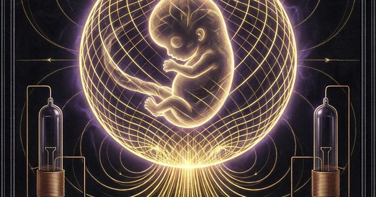
Harold Saxton Burr: L-Fields & Electrodynamic Blueprints
Executive Summary & Theoretical Thesis: Biological Morphogenesis via Macroscopic Field Vectors
The Inadequacy of Localized Biochemical Determinism
Classical molecular biology treats morphogenesis as an emergent epiphenomenon resulting from localized chemical cascades and genetic transcription networks. In this standard paradigm, tissue patterning is governed by reaction-diffusion architectures, such as Turing mechanisms, where local activation and long-range inhibition drive the spatial distribution of diffusible ligands. However, these diffusion-limited models exhibit severe explanatory deficits when tasked with accounting for long-range spatial coherence, instantaneous global coordination across macroscopic biological tissue, and the rapid restoration of anatomical architecture following catastrophic trauma. Molecular diffusion rates in viscous intracellular and extracellular matrices are constrained by Brownian kinetics and steric hindrance; they scale with the square root of time ($\langle x^2 \rangle = 2Dt$), rendering purely stochastic chemical diffusion fundamentally incapable of coordinating millimetric or centimetric organizational events across complex embryonic volumes within short developmental intervals.
Furthermore, reaction-diffusion dynamics suffer from extreme sensitivity to thermal fluctuations and local mechanical perturbations. A biological system relying exclusively on localized biochemical gradients would routinely succumb to morphogenetic noise, yielding disordered phenotypic variations rather than invariant, stereotypic structural plans. The classical framework provides no metric for explaining how detached cells recognize their precise spatial coordinates relative to the whole anatomical topology, nor how complex morphogenetic movements—such as the epiboly and invagination witnessed during amphibian gastrulation—are coordinated with topological fidelity. This explanatory vacuum necessitates an overarching physical mechanism capable of generating macroscopic boundary constraints that precede and organize downstream molecular events.
The structural resilience of living systems demands a global ordering parameter operating at spatial scales far exceeding the Debye screening length of individual biochemical ions. The prevailing genetic-reductionist stance conflates the material substrate of organic forms—the structural proteins, enzymes, and nucleic acid sequences—with the coordinating architectural force that directs their spatial deposition. This monograph establishes that physical biological architecture is governed by macroscopic electrodynamic fields that operate as non-local boundary conditions, imposing spatial constraints upon cellular differentiation, directional migration, and volumetric boundary formation.
Definition of the Life-Field (L-Field) Vector Formalism
Formulated during his tenure at the Yale School of Medicine, the macroscopic electrodynamic field hypothesis advanced by Harold Saxton Burr defines the “Life-Field” (L-field) not as an esoteric or non-physical vital force, but as an empirically measurable, steady-state electrodynamic spatial vector continuum. The L-field is characterized as a macroscopic electrical scalar-potential gradient mapped across the anatomical axes of living systems, generating an anisotropic vector field:
$$\mathbf{E} = -\nabla \Phi$$
where $\Phi$ denotes the spatial distribution of electrodynamic scalar potential established by macroscopic charge separations, non-ohmic ionic currents, and dielectric interfaces within the biological organism.
Across a continuous biological tissue phase bathed in an aqueous electrolyte, the spatial distribution of the macroscopic bioelectric scalar potential $\Phi(\mathbf{r})$ is governed by the non-linear Poisson-Boltzmann formulation:
$$\nabla \cdot \left[ \varepsilon(\mathbf{r}) \nabla \Phi(\mathbf{r}) \right] = -\rho_{\text{macro}}(\mathbf{r}) - \sum_{i} z_i e c_{i,\infty} \exp\left( -\frac{z_i e \Phi(\mathbf{r})}{k_B T} \right)$$
where $\varepsilon(\mathbf{r})$ represents the spatially dependent dielectric permittivity tensor of the cellular matrix, $\rho_{\text{macro}}(\mathbf{r})$ characterizes the immobilized structural charge density associated with the cytoskeleton and fixed extracellular proteoglycan lattices, $z_i$ is the valence of mobile ionic species $i$, $c_{i,\infty}$ is the bulk electrolyte concentration, $e$ is the elementary charge, $k_B$ is the Boltzmann constant, and $T$ is absolute temperature. The boundary conditions are determined by the macroscopic anatomical geometry, yielding a stable spatial coordinate matrix that directs morphogenetic transport.
Within Burr’s framework, the L-field constitutes an operational morphogenetic-field. It provides invariant spatial coordinates that determine cellular orientation, polarity, and mitotic spindle alignment prior to overt histological differentiation. The L-field is distinct from transient, high-frequency oscillatory phenomena like the neurogenic action-potential; it is an enduring, quasi-DC electrodynamic matrix that permeates intra- and extra-cellular domains, governing the anatomical trajectory of the organism throughout its lifespan.
Thermodynamic Non-Equilibrium and Spatial Organization
Biological organisms exist exclusively as open, highly organized non-equilibrium thermodynamic entities. According to Prigogine’s principles of dissipative structures, the maintenance of low internal entropy requires an uninterrupted throughput of free energy derived from metabolic processes. In Burr’s electrodynamic formulation of biology, this thermodynamic throughput is coupled directly to the production and maintenance of macroscopic electromagnetic boundary conditions. The continuous hydrolytic breakdown of adenosine triphosphate (ATP) by electrogenic membrane-bound translocases drives vast ionic concentration disparities across cellular interfaces, transforming the cellular collective into an integrated network of dipolar macroscopic emitters.
These macroscopic electrodynamic boundaries act as macroscopic constraints in the phase space of morphological development. Without the continuous generation of the L-field gradient, metabolic processes would degrade into thermodynamic equilibrium, resulting in thermal death, cellular desynchronization, or unconstrained neoplastic growth. By maintaining a constant dielectric-field and steady-state electrostatic displacement, the organism converts local scalar metabolic energy into long-range vector forces.
This electrodynamic stabilization establishes what Burr identified as the physical blueprint of the organism: an active, homeostatically regulated electrodynamic field vector that continuously preserves the anatomical blueprint against the entropic decay inherent to fluctuating thermodynamic microenvironments.
Historical Lineage & Experimental Precedents: The Yale Instrumentation Revolution
Burr-Northrop Theoretical Synthesis (1935)
The formal conceptualization of the electrodynamic theory of life materialized through an interdisciplinary collaboration at Yale University between Harold Saxton Burr, an experimental neuroanatomist, and Filmer S. C. Northrop, a philosopher of science specializing in the epistemological foundations of modern physics. In their seminal 1935 publication in The Quarterly Review of Biology, “The Electro-Dynamic Theory of Life,” Burr and Northrop addressed the fundamental epistemological division between mechanistic atomism and metaphysical vitalism. They argued that both paradigms failed to account for biological organization: atomism failed to explain the enduring teleological pattern of the organism amid continuous chemical exchange, while vitalism invoked unquantifiable, non-physical agents.
Burr and Northrop synthesized a radical alternative: biological organization is physical, metric, and non-corpuscular, grounded in the field physics of Michael Faraday, James Clerk Maxwell, and Albert Einstein. They argued that the ultimate constituents of organisms are not discrete chemical particles acting purely locally, but a continuous electrodynamic field that organizes these material constituents. Chemical constituents—proteins, carbohydrates, lipids, water, and electrolytes—flow continuously through this electrodynamic field, assuming spatial positions dictated by the field’s topological gradients. By shifting the foundational ontological unit of biology from the particle to the field, Burr and Northrop reconciled biological organization with theoretical physics, asserting that life is governed by electrodynamic field geometry rather than by linear, stochastic chemical encounters.
The Ultra-High Impedance Vacuum-Tube Microvoltmeter
Empirical verification of Burr’s theoretical claims required the resolution of a foundational instrumentation problem. During the 1930s, standard measuring instruments—such as low-resistance D’Arsonval galvanometers—depended on the extraction of electrical current from the system under observation to actuate mechanical measurement indicators. When applied to living tissues, these primitive instruments produced catastrophic electrical distortion. The drawing of current across living membranes induced rapid electrical polarization, altered endogenous potential distributions, elicited neurovascular responses, and generated spurious galvanic artifacts.
+-------------------------------------------------------------------------+
|                  BURR-LANE ELECTROMETER SCHEMATIC                       |
|                                                                         |
| Biological  [Ag/AgCl]   Saline    Grid     Vacuum Tube     Balanced     |
| Tissue   --> [Electrode] Bridge -> (High) -> (10^12 Ohm) -> Galvanometer|
| Specimen    [Electrode] Bridge             Input Stage     Bridge (0 A) |
|                                                                         |
| Non-Faradaic Boundary: Microvolt Resolution with Zero Polarization      |
+-------------------------------------------------------------------------+
To circumvent this limitation, Burr collaborated with the Yale University low-temperature physicist Cecil T. Lane to design an electrometer circuit tailored for biological systems. Their instrument, an ultra-high input impedance vacuum-tube microvoltmeter, utilized specialized electrometer vacuum tubes with an extraordinary input impedance approaching $10^{12}$ ohms ($1,\text{T}\Omega$). The grid circuit of the input stage drew virtually zero current—less than $10^{-12}$ amperes—from the biological tissue under examination.
Primary source documentation: Burr, H. S., Lane, C. T., & Nims, L. F. (1936). A Vacuum Tube Microvoltmeter for the Measurement of Bio-electric Phenomena. Yale Journal of Biology and Medicine, 9(1), 65–76.
- Input Impedance: Approaching $10^{12},\Omega$, preventing biological source loading.
- Grid Current: $< 10^{-12},\text{A}$, avoiding perturbation of endogenous polarization layers.
- Voltage Sensitivity: Continuous resolution down to $1.0,\mu\text{V}$ direct current across high-resistance tissue structures.
- Bridge Configuration: Symmetrical dual-triode balanced Wheatstone-bridge typology running off isolated direct-current storage cells to eliminate alternating-current mains hum and harmonic contamination.
By operating in a true null-deflection and non-loading electrostatic sensing regime, this apparatus permitted, for the first time in experimental embryology and neuroanatomy, the continuous, long-term mapping of endogenous electrodynamic scalar potentials in vivo without destabilizing delicate physiological boundaries.
Mitigation of Electrode Polarization Artifacts
Even with an electrometer drawing negligible current, Burr faced the challenge of electrochemical artifacts generated at the metal-electrolyte interface. When bare metallic electrodes contact biological tissues containing aqueous electrolytes, electrochemical half-cell reactions immediately develop, creating ill-defined electrochemical potentials that obscure endogenous bioelectric field values. Furthermore, mechanical agitation of metallic electrodes causes unpredictable fluctuations in these junction potentials, producing false readings that mimic biological voltage variations.
Burr solved this interface problem through the engineering of non-polarizable, reversible silver/silver chloride ($\text{Ag/AgCl}$) electrodes paired with physiological saline bridges. Pure silver pins were chlorided via electrolysis to establish a stable, reversible thermodynamic equilibrium:
$$\text{Ag} + \text{Cl}^- \rightleftharpoons \text{AgCl} + e^-$$
These reversible elements were housed in glass jackets and physically separated from direct tissue contact through liquid junctions containing isotonic physiological saline.
The biological tissue touched only the chemically inert, non-reactive physiological saline solution. The interface between the tissue and the measuring apparatus was thus converted into a non-polarizable boundary characterized by minimal contact overpotential and an absence of mechanical shearing artifacts. Burr validated these non-polarizable assemblies by systematically rotating the probes, reversing their physical orientation, interchanging electrode leads, and measuring inert chemical substrates, confirming that the recorded microvolt differentials were authentic, endogenous potential gradients of the living organism and not artifacts of the recording apparatus.
Mathematical Formalism & Physical Mechanics: Maxwellian Electrodynamics in Tissue Continua
Maxwellian Steady-State Potential Formulations in Living Systems
The biological tissue continuum operates within a classical macroscopic electrodynamic regime governed by Maxwell’s equations. Given the low-frequency character of the steady-state L-field, the inductive time-derivative term of the magnetic field ($\partial \mathbf{B}/\partial t$) vanishes, reducing the electrodynamic system to an electrostatic and stationary-current configuration:
$$\nabla \times \mathbf{E} = 0 \implies \mathbf{E} = -\nabla \Phi$$
$$\nabla \cdot \mathbf{D} = \rho_f$$
where $\mathbf{D} = \hat{\varepsilon} \mathbf{E}$ represents the electric displacement field, $\hat{\varepsilon}$ is the biological dielectric permittivity tensor, and $\rho_f$ is the local free charge density within the interstitial and cellular compartments.
ANISOTROPIC TISSUE MATRIX
               +-----------------------------------------+
               | Membrane Dielectric   Capacitive L-Field|
               | [======Lipid Bilayer (\epsilon_r \approx 2)======] |
  Metabolic    |                                         | Macro-Scale
  Ion Pumping  |            Intracellular Fluid          | Potential
  (ATP-Driven) |    (\epsilon_r \approx 80, \sigma \approx 1 S/m)| Gradient
               |                                         | \nabla \Phi
               | [======Lipid Bilayer (\epsilon_r \approx 2)======] |
               | Extracellular Matrix (Anisotropic Paths)|
               +-----------------------------------------+
Because living biological matrices are densely packed with semipermeable lipid membranes, aqueous electrolyte fluids, fixed macromolecular proteoglycan charges, and anisotropic collagenous fascia, the current density $\mathbf{J}$ contains both conductive ohmic components and non-ohmic steady-state displacement fluxes. Conservation of continuous electric charge dictates:
$$\nabla \cdot \mathbf{J} = -\frac{\partial \rho_f}{\partial t}$$
Under steady-state physiological operations ($\partial \rho_f / \partial t = 0$), the continuity relation simplifies to:
$$\nabla \cdot \left( \hat{\sigma} \nabla \Phi \right) = 0$$
where $\hat{\sigma}$ is the anisotropic conductivity tensor of the biological tissue. Biological morphology maps directly to this equation: the directional axes of anatomical structures (cephalocaudal, dorsoventral, left-right) are defined by spatial variations in the conductivity tensor $\hat{\sigma}(\mathbf{r})$ and local electrogenic current sources ($I_{\text{source}}(\mathbf{r})$).
The spatial gradient of the scalar potential ($\nabla \Phi$) acts as a vector field that defines the morphology of the tissue continuum. Cells residing within this vector field experience continuous, deterministic electrophoretic and electroosmotic forces that coordinate their structural orientation.
Dielectric Permittivity and Ionic Dipole Moments
Biological tissues are not simple aqueous conductors; they possess extraordinary dielectric-field characteristics across low-frequency ranges. The cellular membrane, a lipid bilayer approximately 5 to 7 nanometers in thickness, presents a massive electrical capacitance on the order of $1.0,\mu\text{F/cm}^2$. This thin dielectric barrier separates two aqueous conducting phases containing physiological ionic species ($\text{K}^+$, $\text{Na}^+$, $\text{Ca}^{2+}$, $\text{Cl}^-$). Consequently, cellular ensembles exhibit an anomalous relative dielectric permittivity ($\varepsilon_r$) that can exceed $10^5$ to $10^6$ in the sub-kilohertz regime due to interfacial Maxwell-Wagner polarization and counterion relaxation phenomena.
This low-frequency dielectric dispersion, designated as the $\alpha$-dispersion region, enables living tissue to store high levels of electrostatic potential energy per unit volume:
$$u_e = \frac{1}{2} \varepsilon_0 \varepsilon_r |\mathbf{E}|^2$$
At this structural interface, macroscopic dipoles align within the structural matrix. Cellular protein structures, particularly the tubulin dimers constituting the microtubular cytoskeleton and the triple-helical tropocollagen fibrils in the extracellular matrix, possess permanent, uncompensated electric dipole moments ($\mathbf{p}$). Under the organizing influence of macroscopic potential gradients ($\nabla \Phi$), these dipolar macromolecules experience mechanical torque:
$$\boldsymbol{\tau} = \mathbf{p} \times \mathbf{E}$$
This electro-mechanical coupling causes alignment along the lines of the electric vector field, transforming the macroscopic L-field into microscopic mechanical strain tensors via converse piezoelectricity and flexoelectricity (such as seen in piezoelectric bone remodeling).
The Continuum Electrodynamic Vector Field Equation
To capture the macro-scale structural stability of the living organism, the electrodynamic field can be modeled as a continuum vector equation where local active biochemical inputs are reconciled with macroscopic spatial boundaries. By combining the conductive, dielectric, and electrogenic contributions of a tissue continuum, we obtain the unified L-field governing equation:
$$\nabla \cdot \left[ \left( \hat{\sigma}(\mathbf{r}) + \frac{\partial \hat{\varepsilon}(\mathbf{r})}{\partial t} \right) \nabla \Phi(\mathbf{r}, t) \right] = \sum_k S_k(\mathbf{r}, t)$$
where $S_k(\mathbf{r}, t)$ represents the spatial distribution of electrogenic metabolic sources, primarily the electrogenic ion-translocating enzymes ($\text{Na}^+/\text{K}^+$-ATPase, V-type $\text{H}^+$-ATPases) that generate transmembrane currents:
$$I_{\text{pump}} = z F J_{\text{pump}}$$
This relationship proves that the L-field is not an ephemeral epiphenomenon, but the direct macroscopic consequence of integrated cellular metabolism acting upon an anisotropic dielectric continuum. When metabolic sources generate spatial potential gradients across gap junctions and extracellular pathways, the resulting macroscopic electric field exerts direct electrophoretic and electroosmotic forces upon charged embryonic morphogens, transmembrane receptor complexes, and cellular surfaces. The L-field is thus a causal physical vector field directing macro-scale biological morphology.
Empirical Evidence & Observational Data: Embryogenesis to Oncological Disruption
Microvoltmeter Mapping of Embryonic Cephalocaudal Axes
Burr provided empirical validation of the L-field hypothesis through longitudinal investigations of embryogenesis in the amphibian Amblystoma punctatum (spotted salamander). Using his non-polarizable microvoltmeter apparatus, Burr (1941) documented macroscopic electrical scalar-potential gradients across the surface of the unfertilized and newly fertilized Amblystoma ovum, long before any histological or morphological evidence of a nervous system or primary embryonic axis had emerged.
Burr, H. S. (1941). Field Properties of the Developing Frog’s Egg. Proceedings of the National Academy of Sciences of the United States of America, 27(6), 276–281. “In the developing egg of Amblystoma, a clear-cut pattern of potential differences exists on the surface of the egg prior to the appearance of any visible morphological structure… The line of maximum potential difference across the egg accurately predicts the future anatomical cephalocaudal axis of the nervous system. The field properties demonstrate an organizing agency whose spatial distribution precedes the overt differentiation of living form.”
Burr immobilized the developing eggs within an isotonic fluid chamber under microscopic visualization, positioning the non-polarizable microvoltmeter electrodes at diametrically opposed coordinates on the surface. Burr detected potential gradients of 10 to 100 microvolts across the ovum. By tracking individual ova through subsequent cleavage, gastrulation, and neurulation, Burr proved that the electrical axis of maximum potential gradient matched the primary cephalocaudal axis of the mature salamander nervous system. The electrical polarity was not caused by the developing nervous system; rather, the anatomical nervous system materialized along the trajectory dictated by the primary electrodynamic field vector.
Pre-Symptomatic Detection of Malignancy and Neoplasms
Burr’s application of the high-impedance microvoltmeter to pathological states yielded empirical discoveries in oncological etiology. If the normal L-field maintains cellular differentiation and tissue boundary conditions, Burr reasoned that oncogenesis must involve a collapse or radical distortion of this electrodynamic boundary architecture.
Working with a genetically defined mouse strain predisposed to spontaneous mammary adenocarcinoma (the Strong A strain), Burr recorded potential gradients across the thoracic and pelvic tissue sectors over several months before any palpable tumor appeared.
       PRE-SYMPTOMATIC ONCOLOGICAL POTENTIAL TRAJECTORY
  Potential
  Shift (mV)
    ^
+50 |                                             /-- Tumor Palpable
    |                                            /    (Histological Malignancy)
+25 |                        /------------------/
    |                       /   Electrodynamic
  0 |                      /    Boundary Inversion
    |  ===================/     (Phase Shift)
-25 |  Healthy Steady-State
    +---------------------------------------------------> Time (Weeks)
       Week 0            Week 10            Week 14       Week 16
In these longitudinal cohorts, Burr observed that animals developing spontaneous mammary tumors exhibited distinct, sustained microvolt potential shifts (frequently reaching 10 to 40 millivolts) across the target anatomical zone up to two to three weeks before the physical appearance of palpable or histologically detectable tumors. The local tissue potential inverted its polarity relative to reference points, signaling a destabilization of normal macroscopic field control.
Subsequent clinical investigations conducted with gynecologists at the Yale School of Medicine demonstrated that female patients harboring malignant cervical or uterine neoplasms exhibited massive direct-current potential shifts across the cervix relative to an abdominal reference site. These shifts were absent in healthy controls or in patients with benign inflammatory conditions. In Burr’s framework, malignancy is primarily an electrodynamic breakdown: a loss of macroscopic field containment that releases the local cellular lineage from its boundary constraints, leading to uncontrolled proliferation.
Diurnal, Lunar, and Solar Electrodynamic Oscillations
To investigate whether biological L-fields operate in isolation or interact with terrestrial and solar electromagnetic environments, Burr initiated a continuous multidecadal recording project. In the late 1930s, he inserted pure silver/silver chloride electrodes directly into the cambium layer of living hardwood trees (primarily Ulmus americana and Acer saccharum) on the grounds of the Yale University campus and in the forests of Old Lyme, Connecticut. These electrodes were interfaced via shielded cables to automatic recording microvoltmeters, generating continuous, uninterrupted DC records spanning more than twenty consecutive years.
The resulting data demonstrated that the potential difference across the cambium of living trees oscillates according to well-defined, multi-frequency cycles. In addition to local temperature and hydration cycles, the voltage traces exhibited periodicities matching:
- Diurnal variations driven by solar atmospheric ionospheric currents ($S_q$ systems);
- Synodic lunar rhythms correlating with gravitational and ionospheric tides;
- Distinct, large-amplitude shifts corresponding to major solar flares and terrestrial geomagnetic storms.
These findings confirmed that biological electrodynamic fields are not closed circuits isolated within individual skins. Organisms operate as open electrodynamic networks coupled to the macroscopic fields of their planetary environment, including Schumann resonances biological coupling. Burr observed that living cambium potentials fluctuated synchronously across trees separated by miles of terrain, demonstrating that large-scale environmental electrodynamic field shifts entrain the endogenous L-fields of terrestrial organisms.
Comparative Paradigms: Morphogenetic Fields in Electrodynamics vs. Molecular Genetics
Electrodynamic L-Fields vs. Chemical Morphogen Cascades
Modern developmental biology relies heavily on models of biochemical morphogen gradients, wherein ligands such as Bicoid, Sonic Hedgehog (Shh), Bone Morphogenetic Proteins (BMP), and Wnt act as concentration-dependent determinants of cellular fate. According to this paradigm, a morphogen source establishes a spatial distribution characterized by monotonic exponential decay:
$$C(x) = C_0 \exp\left(-\frac{x}{\lambda}\right)$$
where $\lambda = \sqrt{D/\kappa}$ represents the characteristic decay length defined by the diffusion coefficient $D$ and degradation rate $\kappa$. Cells measure this local concentration via cell-surface receptors and activate specific transcriptional profiles in response to definite biochemical thresholds.
While chemical gradients participate in local cellular differentiation, they do not resolve the primary spatial coordination problem of embryogenesis. Chemical diffusion across embryonic volumes is slow and subject to variable transport delays, structural degradation, and turbulent mechanical convection. In contrast, Burr’s electrodynamic L-field model operates via Maxwellian field propagation:
$$\mathbf{E}(\mathbf{r}, t) = -\nabla \Phi(\mathbf{r}, t) - \frac{\partial \mathbf{A}(\mathbf{r}, t)}{\partial t}$$
which establishes spatial field patterns across tissues near the speed of light in the dielectric medium. The L-field generates instantaneous physical coordinate axes, organizing charged ligands and receptor complexes via electrophoresis along the tissue membrane:
$$v_{\text{drift}} = \mu_e \mathbf{E}$$
where $\mu_e$ is the electrophoretic mobility of the molecular complex. Rather than relying on Brownian diffusion to construct anatomical forms, the living organism utilizes its electrodynamic field vector to position morphogenetic molecules. In this model, chemical morphogens are structural effectors, while the macroscopic L-field provides the underlying organizational vector field.
Burr Electrodynamic L-Field Model
- Physical Carrier: Macroscopic quasi-DC electric scalar potentials ($\Phi$) and dynamic dielectric displacement fields.
- Measurement: Quantified directly using SI units ($\mu\text{V}$, $\text{mV}$) via ultra-high input impedance ($>10^{12},\Omega$) electrometers.
- Causal Vector: Macroscopic boundary conditions direct local cellular polarization, spindle alignment, and gene expression.
- Pathology: Neoplasia begins as a localized breakdown of field potential architecture, preceding histological aberrations.
Turing-Gradient Molecular Model
- Physical Carrier: Diffusible macromolecular peptide ligands (BMP, Shh, Wnt) moving through Brownian diffusion.
- Measurement: Destructive spatial transcriptomics, fluorophore tag assays, and localized Western blot assays.
- Causal Vector: Local molecular signaling cascades generate macroscopic tissue structures as an emergent epiphenomenon.
- Pathology: Neoplasia is driven by stochastic genetic mutations within oncogenes and tumor-suppressor sequences.
Burr’s Field Vector vs. Sheldrake’s Morphic Resonance
Burr’s L-field theory must be distinguished from the later concept of “morphic resonance” formulated by Rupert Sheldrake in his hypothesis of formative causation. Sheldrake proposed that morphogenetic fields operate across space and time through non-energetic, non-physical resonance based on phylogenetic memory. While Sheldrake’s concept aims to solve the same foundational problem—how organic form is coordinated across space—it posits a mechanism that operates outside the measurable bounds of classical and quantum electrodynamics, invoking non-material telepathic-like resonance mechanisms that lack physical carrier waves, metric field tensions, or direct laboratory quantifiability.
Burr’s theoretical structure remains grounded in empirical physics. The L-field requires no appeal to non-energetic domains, non-physical interactions, or speculative metaphysics. It consists of physical scalar potentials, continuous dielectric currents, and measurable electromagnetic vector fields. Burr insisted on physical quantifiability: if a field exists, it must exert forces on matter, store potential energy, and be measurable in SI units via non-perturbative physical instrumentation. Burr’s model aligns with orthodox continuum mechanics and Maxwellian electrodynamics. It is an empirically verifiable biophysical construct rather than a non-energetic metaphysical postulation.
Contemporary Resurgence: Levin’s Bioelectric Code
Decades after Burr’s tenure at Yale, the conceptual framework of electrodynamic biological organization experienced a major laboratory resurgence through the work of Michael Levin and his laboratory at Tufts University. Levin’s paradigm of the bioelectric code confirms the mechanics of Burr’s earlier model. Using voltage-sensitive fluorescent reporter dyes (such as DiBAC and CC2-DMPE), microelectrodes, and optogenetics, Levin has demonstrated that steady-state transmembrane potentials ($V_{\text{mem}}$) across cellular collectives form continuous bioelectric spatial patterns that orchestrate anatomical morphogenesis, organ size determination, left-right asymmetry, and limb regeneration.
Levin’s work has established that artificial alteration of endogenous bioelectric gradients directly overrides genomic cues. By pharmacologically or optogenetically depolarizing or hyperpolarizing discrete cellular cohorts, his team has induced the de novo growth of structurally complete, functioning eyes on the gut endoderm of Xenopus embryos, directed ectopic brain structures, and induced complete multi-tissue limb regeneration in typically non-regenerative adult organisms.
Levin’s bioelectric state represents the cellular and molecular implementation of Burr’s L-field. Where Burr identified the macroscopic field vectors using early electrometer vacuum tubes, contemporary bioelectricity tracks the same spatial gradients using high-resolution optical physiology and ion channel pharmacology. This research demonstrates that the macroscopic bioelectric field directs downstream epigenetic expression, confirming Burr’s foundational premise: electrodynamic fields direct the morphological deposition of matter.
Metaphysical Implications & Unified Synthesis: Biological Organization as Field Geometry
Living Organisms as Macroscopic Waveguide Systems
Synthesizing Burr’s findings with continuum physical mechanics reveals that the anatomical architecture of complex biological organisms functions as a macroscopic electromagnetic waveguide and resonant cavity. The biological matrix is an anisotropic, multi-layered dielectric medium. It consists of alternating hydrated crystalline phases, structural lipid bilayers, periodic collagen matrices, and highly organized water channels that function as optical, dielectric, and piezoelectric transmission networks.
Within this architecture, the L-field serves as a standing electrodynamic wave that provides spatial stabilization. Chemical macromolecules flow through this structural framework: cells continuously replace their proteins, alter their lipid composition, and exchange their fluids, yet the macroscopic geometry of the organism persists throughout its life cycle.
The living form behaves like an electrodynamic standing wave or a hydraulic vortex: while the matter composing the structure is in continuous flux, the organizational geometry of the field remains invariant. Biological form is not primarily localized within the material components, but within the boundary geometry of the electromagnetic field that traps, directs, and configures those components.
Burr’s macroscopic scalar potentials align closely with the physics of interfacial biological water formulated by Giuliano Preparata, Emilio Del Giudice, and Gerald Pollack. When interfacial water contacts hydrophilic biopolymer surfaces (such as the extracellular matrix or cytoskeletal tubulin), it forms macroscopic exclusion zones (EZ water) extending across tens to hundreds of micrometers. These zones are characterized by an organized crystalline lattice, a net negative electrical charge, and an exclusion of mobile solutes, offset by a corresponding cloud of concentrated hydronium ions ($\text{H}_3\text{O}^+$):
$$\left[ \text{H}2\text{O} \right]{\text{bulk}} \xrightarrow{\text{Hydrophilic Interface}} \left[ \text{H}3\text{O}2^- \right]{\text{EZ}} + \left[ \text{H}^+ \right]{\text{mobile}}$$
This macroscopic charge separation functions as a biological liquid-state capacitor, generating steady-state scalar potentials ($\Phi$) that remain stable in the absence of metabolic active transport. Del Giudice and Preparata demonstrated using Quantum Electrodynamics (QED) that biological water forms coherent domains oscillating in phase with an endogenous electromagnetic field. The Burr L-field corresponds physically to the macroscopic envelope of these coherent domains, which couples metabolic thermodynamic dissipation directly to long-range field coherence across the living tissue volume.
Integration with Earth’s Global Electrodynamic Environment
Burr’s multidecadal botanical investigations demonstrate that the biological L-field does not terminate at the organism’s skin. The biological matrix is coupled to the planetary electrodynamic environment. The Earth’s surface and the lower ionosphere constitute a spherical waveguide characterized by low-frequency electromagnetic resonances—the Schumann resonances—driven by global lightning discharges:
$$f_n \approx \frac{c}{2\pi R_E} \sqrt{n(n+1)}$$
yielding fundamental standing frequencies at approximately 7.83, 14.3, 20.8, 27.3, and 33.8 Hz.
Organisms evolved entirely within this background electromagnetic environment. Burr’s cambium potential records demonstrated direct coupling with solar wind fluctuations, geomagnetic storms, and ionospheric perturbations, indicating that biological L-fields exchange energy and information with these planetary fields. The biological organism is not an isolated mechanical entity, but a dynamic node integrated into a global electrodynamic continuum.
This coupling provides external temporal cues that stabilize internal circadian and infradian physiological oscillations. Organisms act as open bioelectric antennas, with endogenous macroscopic L-fields tuned to the fluctuating electromagnetic oscillations of the planetary environment.
The Transcendent Matrix: Matter Conforming to Energy
The work of Harold Saxton Burr establishes an inversion of mechanistic biology. In the Cartesian-Newtonian framework that still underpins conventional biomedicine, matter is foundational: complex structural configurations of matter generate forces, energy fields, and consciousness as emergent epiphenomena. Burr’s electrodynamic theory of life, validated through decades of rigorous empirical measurement, inverts this hierarchy: field energy is foundational, and physical matter conforms to the geometry of the field.
As Burr wrote in his 1972 summary Blueprint for Immortality, the electrodynamic field holds structural molecules in spatial alignment much as an invisible magnetic field organizes iron filings scattered on a sheet of paper. When the magnetic field changes its topology, the filings follow; if the filings are swept away, new filings deposited in the field immediately reproduce the original pattern.
In the biological organism, physical cells, enzymes, nucleic acids, and structural polymers are the material filings. The L-field is the enduring electrodynamic blueprint that directs their spatial deposition, monitors their functional integration, orchestrates their developmental repair, and signals their oncological breakdown. In the living continuum, form is an electrodynamic geometry.
Frequently Asked Questions: Technical and Conceptual Inquiries
Physics of Bioelectric Distinction
How does Burr’s L-field differ fundamentally from conventional diagnostic bioelectric signals like the ECG, EMG, and EEG?
Standard diagnostic signals—such as the electrocardiogram (ECG), electromyogram (EMG), and electroencephalogram (EEG)—capture transient, alternating-current (AC) electrical potentials generated by the synchronized firing of action potentials across excitable membranes (e.g., cardiomyocytes, skeletal muscle fibers, cortical pyramidal neurons). These signals are high-frequency, time-varying oscillatory phenomena (typically 0.5 Hz to 500 Hz) that reflect transient changes in membrane permeability driven by voltage-gated $\text{Na}^+$ and $\text{K}^+$ channels. Once the depolarization-repolarization wave passes, the local electrical differential returns to baseline.
Burr’s L-field, conversely, is an enduring quasi-DC (direct current) macroscopic potential gradient. It does not depend on excitable neural or muscular membranes and is present across non-excitable tissues, including plant cambium, epithelial sheets, and unfertilized embryonic ova. Where the EEG and ECG measure transient functional messages propagating through an already structured biological network, the L-field measures the underlying, steady-state electrodynamic spatial framework that organizes the tissue substrate itself. The L-field is a structural and morphogenetic field, not a transient physiological signal.
Artifacts vs. Endogenous Potentials
Did Harold Saxton Burr measure authentic endogenous biological potentials, or were his microvoltmeter readings artifacts of contact potentials, galvanic skin responses, and movement dynamics?
This was the central methodological challenge addressed by Burr throughout his career at the Yale School of Medicine. Conventional low-impedance galvanometers inevitably generate electrochemical artifacts: they draw electrical current, which polarizes tissue interfaces, creates non-Faradaic overpotentials, and induces galvanic chemical reactions that reflect the composition of the electrodes rather than the state of the tissue.
Burr avoided these artifacts through two technical implementations. First, with Cecil T. Lane, he developed a custom vacuum-tube microvoltmeter operating with an input impedance approaching $10^{12},\Omega$. The instrument measured scalar potentials with near-zero current draw ($<10^{-12},\text{A}$), preventing tissue polarization. Second, he eliminated metal-to-tissue contact by using reversible, non-polarizable $\text{Ag/AgCl}$ electrodes suspended in isotonic physiological saline bridges. Burr confirmed that the observed microvolt potentials were authentic biological properties through systematic controls: swapping electrode poles, rotating the orientation of specimens relative to the probes, measuring non-living controls, and tracking consistent voltage patterns that mapped predictably to embryonic development and oncological transformations.
+--------------------------------------------------------------------------+
|          INSTRUMENTAL CIRCUIT DESIGN COMPARISON                          |
|                                                                          |
| A. Conventional Galvanometer (Artifact-Prone):                           |
|    Tissue Source (R_in) --> Low Z Meter (~10^3 Ohm)                      |
|    Result: High current extraction -> Tissue polarization -> Artifacts   |
|                                                                          |
| B. Burr-Lane Electrometer (Artifact-Free):                               |
|    Tissue Source (R_in) --> Ag/AgCl / Saline Bridge --> Electrometer Grid|
|                             (Input Z ~ 10^12 Ohm)                        |
|    Result: Zero current extraction (<10^-12 A) -> Pure Endogenous L-Field|
+--------------------------------------------------------------------------+
Contemporary Clinical Relevance
How does Burr’s L-field framework relate to modern clinical technologies, such as pulsed electromagnetic field (PEMF) therapies and bioelectric regeneration?
Burr’s foundational work directly anticipates modern electroceuticals and regenerative bioelectricity. In orthopedics, the understanding of electrodynamic fields inspired the development of FDA-approved pulsed electromagnetic field (PEMF) devices used to treat recalcitrant non-union bone fractures. These devices operate by inducing localized electric vector fields that stimulate osteoblastogenesis via converse piezoelectric pathways and voltage-sensitive calcium channels, matching Burr’s model of electrodynamic architectural control.
In regenerative medicine, Burr’s insights have been confirmed by contemporary bioengineering laboratories, such as Michael Levin’s group at Tufts University. Researchers now target bioelectric gradients to induce structural regeneration in complex organs, restore neural patterns after brain defects, and normalize malignant phenotypes without toxic chemotherapy. Burr’s discovery that malignant transformations are preceded by distinct microvolt potential shifts has re-emerged in early cancer detection methodologies that identify disrupted bioelectric epithelia. Contemporary biophysics continues to validate Burr’s primary finding: targeted modulation of the macroscopic bioelectric blueprint can orchestrate complex, multi-scale biological outcomes.