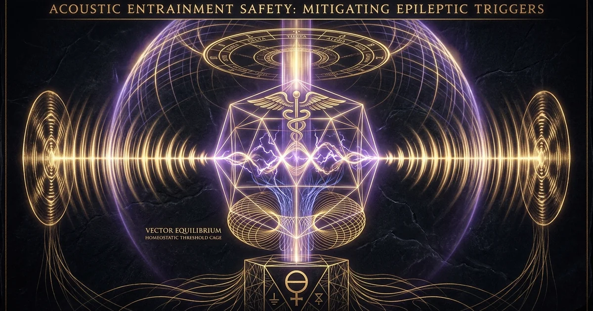
Acoustic Entrainment Safety: Mitigating Epileptic Triggers
Protocol Overview & Neurophysiological Thesis
Acoustic neuromodulation, achieved predominantly through dichotic auditory beat stimulation, relies on the biological computation of phantom phase-differentials within the brainstem. Rather than driving primary neocortical circuits through sheer sensory overdrive, this modality exploits microsecond interaural time differences to elicit subcortical phase-locking. However, modern clinical applications often conflate auditory driving mechanics with visually driven photoparoxysmal cascades. A precise physiological delineation reveals that while the primary visual cortex can be forced into catastrophic, high-amplitude hypersynchrony through stroboscopic light, auditory driving operates under far more subtle biophysical constraints. The critical vulnerability emerges not from standard auditory frequency following alone, but from the unmonitored intersection of subcortical driving with unstable thalamocortical networks, latent channelopathies, and multimodal sensory summation.
The clinical objective of neuro-entrainment protocols is the targeted shift of baseline neural oscillations into functional states of relaxation, heightened vigilance, or transpersonal integration. Yet, altering baseline cortical excitability carries intrinsic hazards when applied to heterogeneous populations. By driving electrophysiological rhythms toward specific target bands—such as Theta (4.0–8.0 Hz) or Gamma (30.0–100.0 Hz)—practitioners deliberately modulate the excitation-inhibition ($E/I$) balance regulated by cortical GABAergic interneurons. In individuals with intact neurochemical homeostasis, this driving facilitates functional hemispheric synchronization and deep meditative absorption. In vulnerable neural topographies, however, shifting the balance of cortical excitability can lower the seizure threshold, destabilizing latent epileptogenic foci that would otherwise remain dormant under standard sensory processing.
To ensure safety across clinical and contemplative applications, practitioners must maintain a rigorous diagnostic boundary separating broad idiopathic epilepsies from specific reflex epileptogenic phenotypes. Idiopathic generalized epilepsies are governed by diffuse genetic channelopathies that predispose thalamocortical loops to spontaneous synchronous oscillations, typically manifesting as generalized spike-and-wave discharges. Conversely, reflex epilepsies require an exogenous trigger—classically stroboscopic flicker, though rarely complex music or precise auditory tones—to breach paroxysmal thresholds. Clinical safety guidelines must distinguish between subcortical phase-locking dynamics and primary sensory seizure generation, ensuring that baseline screening isolates both generalized susceptibility and narrow sensory sensitivities before initiating sound-based driving protocols.
“Photic- and pattern-induced seizures are defined electroencephalographically by the photoparoxysmal response (PPR), an abnormal cortical response characterized by generalized or localized spike-and-wave discharges evoked by intermittent photic stimulation.”
— Fisher, R. S., et al. (2005). Photic- and pattern-induced seizures: a review for the Epilepsy Foundation of America Working Group. Epilepsia, 46(9), 1426-1441.
“Auditory beat stimulation, whether binaural or monaural, modulates cortical electrophysiology through subcortical phase-locking within the superior olivary complex. However, the resulting frequency-following response remains biologically distinct from cortical hypersynchrony, exhibiting significantly lower amplitudes than photic driving responses and possessing negligible intrinsic paroxysmal capacity in healthy human subjects.”
— Chaieb, L., et al. (2015). Auditory Beat Stimulation and its Effects on Cognition and Electrophysiological Brain Waves. Frontiers in Psychiatry, 6, 70.
Multimodal stimulation paradigms, specifically those coupling acoustic binaural stimulation with rhythmic luminance modulation, amplify paroxysmal risk exponentially. When auditory pulses coincide with periodic optical flicker within the vulnerable 15–25 Hz window, the central nervous system experiences sensory summation across independent ascending pathways. The visual system’s direct, retinogeniculo-striate projection directly activates primary visual cortex pyramidal neurons, while the ascending auditory pathway delivers synchronized discharges via the medial geniculate body to the temporal cortex. If these orthogonal sensory streams converge upon an excitable parietal-occipital network, the resulting spatial and temporal summation can instantly overcome GABAergic inhibition. Thus, clinical safety guidelines mandate the total decoupling of acoustic entrainment from photic driving whenever an individual presents with ambiguous neurological histories, familial seizure markers, or undetermined neurological contraindications.
Acoustic Entrainment vs. Photic Hyperexcitability: Mechanistic Boundaries
The physiological divergence between auditory and photic entrainment rests upon the neuroanatomical architecture of their respective sensory transductive pathways. Visual entrainment relies upon the retina’s direct phototransduction cascade, which routes rapid, high-luminance impulses through the optic nerve directly to the lateral geniculate nucleus (LGN) of the thalamus, and subsequently to layer IV of the primary visual cortex (V1). This pathway is uniquely vulnerable to the photoparoxysmal-response (PPR), wherein high-contrast, intermittent stroboscopic light bypasses local inhibitory interneuronal gates. The resulting massive synchronization across millions of contiguous occipital pyramidal neurons can trigger a paroxysmal depolarizing shift (PDS), manifesting as generalized epileptiform discharges. The physical amplitude of photic cortical evoked potentials is exceptionally high, making visual driving one of the most potent triggers of reflex seizures known to clinical neurology.
In stark contrast, auditory driving via frequency-following-response mechanics does not originate within the neocortex. Dichotic auditory beats—frequently termed binaural-beats—are generated entirely by subcortical central processing. When two continuous sinusoidal tones of slightly differing frequencies are introduced separately to each ear, the mechanical movement of the basilar membrane within the cochlea transposes these frequencies into discrete phase-locked action potentials along the auditory nerve (cranial nerve VIII). These distinct neural spike trains ascend directly to the brainstem, converging upon the superior olivary complex. The phase difference between the two carrier frequencies is processed via subcortical dendritic delay lines, producing a centrally perceived amplitude modulation. The amplitude of this acoustic frequency-following response (FFR) measured at the scalp via electroencephalography (EEG) is remarkably subtle—typically quantified in tenths of a microvolt ($\mu\text{V}$)—in sharp contrast to the massive hundreds-of-microvolt paroxysmal spikes provoked during photic driving.
Consequently, the seizure risk acoustic entrainment poses must not be conflated with the robust, proven seizure-inducing capacity of intermittent photic stimulation. Auditory beat stimulation lacks the direct, uninhibited thalamocortical drive inherent to the visual pathway; it operates as an endogenous phase-modulation rather than an exogenous sensory flash. Nevertheless, the systemic vulnerability resides in the potential for this subtle brainstem-mediated modulation to resonate with an already destabilized neocortical architecture. While pure acoustic entrainment possesses a vanishingly small capacity to initiate primary de novo epileptogenesis in a structurally intact brain, its ability to perturb cortical synchronization requires that it be treated with strict clinical caution in populations prone to hyperexcitability.
Target Consciousness States and Cortical Excitability Baselines
Acoustic neuromodulation is primarily deployed to guide the global electroencephalographic profile into specific brainwave frequencies to cultivate altered states of consciousness, optimize neuroplasticity, or facilitate transpersonal introspection. In the context of the Monroe Gateway Protocol analysis, precise frequency manipulation is intended to induce a condition known as hemispheric-synchronization, wherein the bioelectrical activity across both cerebral hemispheres demonstrates coherent phase relationships. These target consciousness states are traditionally categorized across canonical spectral bands:
Delta (0.5 – 4.0 Hz): Profound somatic repair, unconscious baseline
Theta (4.0 – 8.0 Hz): Hypnagogia, deep transpersonal imagery, limbic access
Alpha (8.0 – 12.0 Hz): Attentive detachment, sensorimotor gating, baseline stabilization
Beta (12.0 – 30.0 Hz): Active cognition, external orientation, executive processing
Gamma (30.0 – 100.0 Hz): Cross-modal binding, hyper-vigilance, peak states
The clinical danger lies in how these frequency transitions alter the fundamental baseline of cortical excitability. When an acoustic entrainment protocol drives a brain toward low-frequency regimes—specifically deep Theta or Delta—the global firing rate of cortical pyramidal cells decreases, accompanied by an increase in burst-firing dynamics regulated by low-threshold calcium channels ($T$-type channels) within the thalamus. In an individual with an intact central nervous system, this promotes the subjective experience of somatic disengagement and visionary hypnagogia. However, in individuals with latent generalized spike-and-wave tendencies—such as those with juvenile absence epilepsy—the natural 3.0–4.0 Hz oscillation of thalamocortical circuits is dangerously close to these target acoustic driving frequencies. An exogenous entrainment protocol sustained at 4.0 Hz can theoretically stabilize the exact rhythmic burst-firing patterns that sustain an absence seizure.
Conversely, driving the brain into high-frequency Beta or high-amplitude Gamma bands increases overall cortical metabolism and demands substantial GABAergic inhibitory activity to prevent runaway recurrent excitation. In individuals possessing subclinical channelopathies affecting voltage-gated sodium or potassium channels (such as variants in the SCN1A or KCNQ2 genes), the sustained entrainment of Gamma frequencies can overwhelm parvalbumin-positive ($PV^+$) GABAergic basket cell networks. Once local inhibitory capacity is saturated, the entrainment shift precipitates a transition from controlled high-frequency binding into localized epileptiform spiking, transforming a session intended to induce cognitive peak performance into an acute neurological crisis.
Clinical Stratification: Idiopathic vs. Reflex Epileptogenic Susceptibility
Establishing clinical safety guidelines for auditory beat stimulation requires precise neuro-diagnostic stratification of patients into distinct susceptibility tiers. Idiopathic generalized epilepsies (IGEs)—encompassing childhood absence epilepsy, juvenile absence epilepsy, juvenile myoclonic epilepsy, and epilepsy with generalized tonic-clonic seizures alone—account for roughly one-third of all epilepsies. These syndromes are characterized electroencephalographically by bilateral, synchronous, symmetrical paroxysmal discharges emerging spontaneously across normal background rhythms. Because the underlying pathology in IGE is a generalized hypersynchrony mediated by intrinsic thalamocortical networks, any external stimulus that promotes broad hemispheric coherence can theoretically act as a permissive environment for seizure propagation, even if the primary sensory trigger is non-paroxysmal.
Reflex epilepsies, by contrast, are conditions where seizures are reliably and consistently precipitated by a specific, external or internal sensory trigger. While photosensitive epilepsy is the most recognized reflex phenotype, affecting approximately 3% of individuals with epilepsy, sound-induced reflex seizures occupy a distinct and complex clinical spectrum. Sound-induced reflex epilepsies are traditionally bifurcated into two categories:
- Simple sound-induced epilepsy (audiogenic/acoustic epilepsy): Direct, sudden, high-intensity auditory shocks (startle-induced seizures) or hyper-specific acoustic frequencies that directly depolarize an excitable focus in Heschl’s gyrus.
- Musicogenic epilepsy: Highly complex, integrated temporal-limbic seizures triggered not merely by the acoustic mechanics of sound, but by the affective, emotional, and mnemonic integration of musical structures over extended exposure periods (typically several minutes).
Clinical stratification demands that practitioners distinguish between an idiopathic patient whose broad seizure threshold might be lowered by prolonged acoustic driving, and a reflex patient whose brain possesses an isolated, frequency-specific trigger. In patients with true reflex musicogenic epilepsy, the trigger is typically linked to the affective and structural dimensions of the auditory stimulus rather than the microsecond phase-differentials characterizing binaural beats. However, because both conditions can present with latent, undiagnosed manifestations, screening protocols must address the shared vulnerability: an intrinsic inability of the central nervous system to attenuate synchronous sensory input through normal GABAergic inhibitory mechanisms.
Biophysical Mechanisms & Brainwave Dynamics
The biophysical pathway through which binaural acoustic stimulation impacts central brainwave activity relies upon central auditory computational neuroanatomy. Unlike monaural beats—which are physical amplitude modulations created by the acoustic interference of two sound waves before entering the ear canal—binaural beats are a purely neurocomputational construct. They are generated when two continuous, pure sinusoidal acoustic waves of differing frequencies, but identical amplitudes, are applied separately to each ear through isolating stereo transducers. The physical sound waves do not interact mechanically in the air or along the tympanic membrane; their integration is an active biological computation executed within the lower brainstem.
The ascending pathway depicted above traces how dichotic signals migrate from mechanical peripheral vibrations to generalized electrophysiological modulations across the neocortical mantle. The critical transformation occurs when the brainstem’s coincidence-detection machinery converts a physical phase-differential into an oscillatory firing rate that ascends via the lateral lemniscus into the midbrain and thalamus. Once the thalamus integrates these rhythmic volleys, the resulting resonance shifts global cortical networks via recurrent thalamocortical loops, which under pathological conditions can degrade into paroxysmal dysrhythmia.
Superior Olivary Complex Mechanics and the Frequency Following Response (FFR)
The physical site of binaural beat creation is the medial superior olive (MSO), an integral nucleus of the superior olivary complex located in the pons. The primary evolutionary purpose of the MSO is horizontal sound localization, mediated through the computation of interaural time differences (ITDs). Neurons within the MSO receive bilateral excitatory glutamatergic inputs from spherical bushy cells located within the anterior ventral cochlear nucleus (AVCN). These bushy cells maintain immaculate phase-locking to the incoming acoustic waveforms, preserving temporal precision down to the microsecond level.
The internal architecture of the MSO functions as a biological delay line, precisely configured to compute spatial disparities through coincidence detection:
$$\tau = \frac{\Delta \theta}{2\pi \cdot f_c}$$
where $\Delta \theta$ represents the interaural phase disparity and $f_c$ denotes the fundamental carrier frequency. As the two pure tones arrive at each ear with a deliberate, fixed phase difference—for example, 200 Hz in the left ear and 208 Hz in the right ear—the interaural phase difference continuously drifts through 360 degrees at a rate exactly equal to the mathematical difference between the two inputs:
$$f_{\text{differential}} = |f_{\text{right}} - f_{\text{left}}| = 8\text{ Hz}$$
The neurons of the MSO systematically fire when the bilateral inputs arrive simultaneously, causing the population firing rate of the MSO to cyclically modulate at this 8 Hz differential.
This synchronized brainstem discharge propagates rostrally via the lateral lemniscus to the central nucleus of the inferior colliculus (CNIC). The CNIC serves as an obligate relay, amplifying the phase-locked discharge before projecting it to the medial geniculate body (MGB) of the thalamus. The thalamus, functioning as the central pacemaker for neocortical rhythms, distributes this periodic modulation to the primary auditory cortex (A1; Brodmann areas 41 and 42) via thalamocortical radiation fibers. This process induces the Frequency Following Response (FFR), wherein wide-scale neocortical assemblies align their endogenous firing rhythms to match the exogenous 8 Hz modulation envelope.
Acoustic Physics: Carrier Frequencies, Offset Differentials, and Auditory Processing
The physiological generation of a binaural beat is constrained by strict biophysical boundaries regarding binaural carrier wave physics. As initially documented in the archival laboratory findings of Oster (1973), the human brain’s capacity to process binaural beats relies entirely on the phase-locking fidelity of the auditory nerve fibers. This phase-locking capacity degrades rapidly above 1000 Hz and becomes completely nonexistent at frequencies exceeding 1500 Hz. For optimal central integration, carrier frequencies must reside below 1000 Hz, with the greatest electrophysiological power and subjectively perceived modulation depth achieved when carrier frequencies are calibrated within the 100 Hz to 500 Hz envelope.
The frequency offset—the differential between the left and right acoustic waves—defines the target entrainment rhythm. For clinical safety, this differential is calibrated to align with specific endogenous brainwave bands. If a carrier frequency is positioned at 250 Hz, the application of 256 Hz to the contralateral ear yields an offset differential of 6 Hz, situated squarely within the Theta band:
Audio Input Left:   250 Hz Sine Wave ──┐
                                       ├─► [MSO Computation] ──► 6 Hz Theta Modulation
Audio Input Right:  256 Hz Sine Wave ──┘
Auditory processing demands that the acoustic waveforms remain clean, uncompressed, and pure sinusoidal profiles. When carrier frequencies exceed 500 Hz, the physical spacing of the human head begins to introduce substantial interaural level differences (ILDs) in addition to interaural time differences (ITDs). The central nervous system’s spatial localization relies predominantly on ITDs below 1000 Hz (processed via the MSO) and ILDs above 1000 Hz (processed via the lateral superior olive, or LSO). Driving the brain via carrier frequencies above 1000 Hz transfers the computational load away from phase-locked coincidence detection into amplitude-attenuation circuits, completely eliminating the neurophysiological basis of the binaural frequency following response.
EEG Spectral Bands and Thalamocortical Dysrhythmia Risks
Cortical entrainment follows distinct spectral bands, each governed by specific microcircuit configurations and intrinsic resonant frequencies within the brain:
| Spectral Band | Frequency Range (Hz) | Primary Biological Correlate | Neurochemical Modulators |
|---|---|---|---|
| Delta | 0.5 – 4.0 Hz | Deep slow-wave sleep, metabolic clearance | Adenosine, low Acetylcholine |
| Theta | 4.0 – 8.0 Hz | Memory consolidation, limbic integration | Acetylcholine, hippocampal GABA |
| Alpha | 8.0 – 12.0 Hz | Sensorimotor gating, idling, internal focus | Thalamic GABA, Serotonin |
| Beta | 12.0 – 30.0 Hz | Cortical alert processing, active focus | Dopamine, Norepinephrine |
| Gamma | 30.0 – 100.0 Hz | Local network binding, sensory synthesis | Glutamate (NMDA/AMPA), fast GABA |
The neurophysiological peril of exogenous acoustic driving surfaces when driving parameters interact pathologically with thalamocortical dynamics. Under healthy conditions, the thalamocortical network transitions seamlessly between desynchronized low-voltage fast activity (waking Beta/Gamma) and synchronized high-voltage slow activity (deep sleep Delta). This rhythm is maintained by recurrent inhibitory feedback loops between thalamic relay neurons and the thalamic reticular nucleus (TRN).
Thalamocortical dysrhythmia (TCD) represents a profound functional breakdown of this pacing system. In TCD, hyperpolarization of thalamic relay neurons de-inactivates low-threshold $T$-type calcium channels, shifting the firing pattern from tonic single-spike mode into low-frequency burst-firing mode. This generates a persistent, pathological low-frequency (4.0–7.0 Hz Theta) core surrounded by an uninhibited ring of high-frequency (Gamma) excitation—a phenomenon known electrophysiologically as the “edge effect.” If an acoustic entrainment protocol drives sustained Theta activity in a brain already predisposed to thalamocortical hyperpolarization, it risks entrenching this dysrhythmia. This state can degrade the signal-to-noise ratio of neocortical processing, lower the seizure threshold, and precipitate temporal-lobe paroxysms or non-convulsive focal seizures characterized by profound subjective dissociation, amnesia, and autonomic collapse.
Differential Diagnostics: Acoustic vs. Photic Paroxysmal Triggers
Clinical safety protocols demand rigorous differential diagnosis to separate the neurobiological vulnerabilities provoked by photic versus acoustic sensory driving. While both sensory modalities converge upon thalamic integration hubs, the speed, amplitude, and spatial distribution of their cortical inputs differ fundamentally. Photosensitivity remains the archetypal reflex trigger because the mammalian neocortex possesses vast evolutionary machinery dedicated to rapid, high-bandwidth optical edge- and motion-detection. This system can be rapidly converted into wide-scale synchronous paroxysms when exposed to high-contrast luminance shifts. Sound, though processed with superior microsecond temporal resolution, distributes its energetic impact across brainstem and midbrain waystations before engaging neocortical assemblies.
Pure Acoustic Binaural Entrainment
- Target Structures: Medial Superior Olive (MSO), Inferior Colliculus, Medial Geniculate Body, Primary Auditory Cortex (A1).
- Paroxysmal Risk: Extremely low in non-epileptic populations; moderate in verified musicogenic or temporal reflex phenotypes.
- Primary Threat Frequencies: Abrupt transitions; resonant match with 3.0 Hz spike-wave absence dynamics or prolonged hyper-Gamma (>40 Hz).
- Population Prevalence: True acoustic reflex epilepsy affects less than 1 in 100,000 individuals within the general population.
Photic Stroboscopic Stimulation
- Target Structures: Retina, Lateral Geniculate Nucleus (LGN), Striate Cortex (V1), Extrastriate Cortices, Intracortical Pyramidal Layers.
- Paroxysmal Risk: Exceptionally high in photosensitive populations; capable of provoking paroxysmal depolarizing shifts (PDS).
- Primary Threat Frequencies: High vulnerability between 15.0 Hz and 25.0 Hz; elevated risk across the broader 10.0–30.0 Hz spectrum.
- Population Prevalence: Photosensitivity occurs in ~3% of all epileptic patients, rising to 10% in juvenile idiopathic phenotypes.
The Strobe Vulnerability Matrix: Critical Flicker-Fusion and Cortical Drive
Photosensitive epilepsy represents the absolute standard for sensory reflex epileptogenesis. The human visual system processes light via photoreceptors that transmit rapidly to bipolar and ganglion cells, which then project to the parvocellular and magnocellular layers of the LGN. When a stroboscopic light source flashes within the visual field, each flash elicits a massive compound visual evoked potential (VEP) within the primary visual cortex. As the flash rate increases, the human visual system approaches the Critical Flicker-Fusion (CFF) threshold—the frequency at which intermittent light flashes fuse into the perception of continuous, steady illumination, typically between 30 Hz and 60 Hz depending on luminance and retinal eccentricity.
The neurophysiological danger zone for photoparoxysmal responses occurs just below the CFF threshold, specifically concentrated within the 15 Hz to 25 Hz window:
Safe Basal Zone:       0.5 Hz ──► 8.0 Hz   (Low cortical driving capacity)
Vulnerability Matrix: 10.0 Hz ──► 14.0 Hz  (Moderate paroxysmal risk)
CRITICAL HAZARD ZONE: 15.0 Hz ──► 25.0 Hz  (MAXIMUM photoparoxysmal / PPR elicitation)
Sub-Fusion Decay:     26.0 Hz ──► 35.0 Hz  (High paroxysmal risk, tapering)
Critical Fusion:      40.0 Hz+             (Approaching perceptual fusion, lower PDS risk)
Within this 15–25 Hz window, the interval between light flashes matches the intrinsic refractory recovery period of neocortical pyramidal neurons and interneurons in V1. Each consecutive flash strikes the visual cortex precisely as its pyramidal cells are recovering from their previous refractory state, compounding post-synaptic excitatory potentials (EPSPs). This rhythmic summation systematically overwhelms local feedforward and feedback GABAergic inhibitory networks, precipitating widespread cortical synchronization that propagates rapidly across corticocortical pathways into the motor cortex and thalamus, precipitating generalized tonic-clonic or myoclonic seizures.
Sensory Summation: The Compounding Danger of Audiovisual Entrainment (AVE)
While pure acoustic entrainment operates with low paroxysmal potential, commercial and clinical modalities frequently deploy Audiovisual Entrainment (AVE) systems. These technologies pair dichotic binaural or monaural beats delivered through headphones with stroboscopic, rhythmic optical pulses delivered via specialized light-emitting diodes (LED) embedded within opaque glasses. The underlying rationale is to leverage multi-sensory binding to enforce rapid, deep brainwave entrainment. However, from a neurophysiological safety perspective, this pairing introduces catastrophic sensory summation risks for susceptible individuals.
When auditory driving pulses are phase-locked directly to optical flashes, two independent, high-capacity sensory pathways deliver synchronized depolarization volleys simultaneously into the central nervous system. The LGN projects through the optic radiations directly to the occipital pole, while the MGB projects via the acoustic radiations to the superior temporal gyrus. Under normal waking conditions, the brain uses cross-modal phase resetting to isolate and process these sensory streams independently without destabilizing cortical equilibrium. Under synchronized AVE driving, these sensory boundaries collapse:
The auditory input acts as a spatial and temporal amplifier for the photic driving signal. EEG recordings during multimodal AVE demonstrate that the presence of an auditory beat synchronized with an optical flicker enhances the amplitude of the visually evoked paroxysmal response by up to 40% compared to optical flicker alone. The combined sensory drive depolarizes vast cortical territories spanning the occipital, parietal, and temporal lobes simultaneously. This widespread multi-modal depolarizing wave bypasses the local inhibitory interneuronal gates that would typically prevent focal paroxysms from spreading, converting a manageable subclinical genetic predisposition into an immediate, severe generalized seizure.
Musicogenic and Sound-Induced Epilepsies: Electroclinical Phenotypes
Sound-induced reflex seizures are rare but clinically vital phenomena that present unique electroclinical phenotypes. In stark contrast to photosensitive epilepsy, which is mediated by primitive, low-level sensory circuits in V1, musicogenic epilepsy represents one of the most cognitively sophisticated reflex seizure disorders recognized by neurology. As established by McDonnell et al. (2007), musicogenic seizures originate predominantly within the non-dominant (typically right) temporal lobe, involving the superior temporal gyrus, the heschl’s gyrus, and deeply integrated limbic and paralimbic structures including the amygdala and hippocampus.
The typical clinical presentation of musicogenic epilepsy does not involve instantaneous paroxysmal spikes upon hearing a pure tone. Instead, it involves an extended, cumulative electroclinical cascade:
Acoustic Exposure (Carrier Waves)
        ──► Affective / Mnemonic Processing (Limbic System)
        ──► Autonomic Hyper-Arousal (Sympathetic Shift)
        ──► Focal Temporal Spike-and-Wave Discharges
        ──► Focal Seizure with Impaired Awareness (Focal Dyscognitive)
The triggers in musicogenic epilepsy are rarely pure acoustic frequencies; they are usually complex melodies, specific vocal timbres, or emotionally resonant musical compositions that evoke intense autonomic and affective processing. However, a subtype of sound-induced reflex epilepsy involves pure-tone audiogenic seizures, wherein exposure to precise carrier frequencies—often between 200 Hz and 500 Hz—initiates focal paroxysms. In these cases, the seizure focus consists of a localized microdysgenesis or cortical dysplasia situated directly within primary auditory cortex A1.
When an individual with undiagnosed audiogenic or musicogenic susceptibility is subjected to prolonged dichotic binaural beats, the continuous, unyielding presentation of the carrier tone can drive this focal hyperexcitable pool of neurons into continuous phase-locked resonance. The focal seizure often manifests subtly: the patient reports sudden epigastric rising sensations, unprovoked terror, auditory distortions (such as an abrupt shift in pitch, hollow echoing, or internal sirens), or micro-myoclonic facial twitching. Without rapid decoupling, these focal seizures can evolve into bilateral tonic-clonic events through secondary generalization.
Step-by-Step Clinical Safety Protocol
The deployment of acoustic neuromodulation protocols requires the systematic application of clinical safety guidelines to eliminate seizure risk and prevent adverse autonomic or psychiatric decompensation. A structured protocol moves progressively through four distinct phases: pre-session neurological risk stratification, precise transducer and carrier calibration, non-linear dynamic frequency modulation, and active post-session baseline stabilization.
[ Phase 1: Screening & Stratification ]
        │
        ▼
[ Phase 2: System Calibration (150–440 Hz) ]
        │
        ▼
[ Phase 3: Linear Ramp Entrainment Protocol ]
        │
        ▼
[ Phase 4: Active Deceleration & Alpha Stabilization (10 Hz) ]
Pre-Session Neurological Screening and Risk Stratification
Before any acoustic driving protocol begins, practitioners must execute a formal, written neurological screening assessment. This screening tool is designed to uncover latent risk factors that categorize a subject into either an absolute contraindication tier (prohibiting protocol initiation) or a relative contraindication tier (demanding frequency adjustments and constant monitoring).
The mandatory screening sequence consists of the following targeted diagnostic assessments:
- Epilepsy and Reflex Seizure History: Explicit evaluation of personal or first-degree familial history of any seizure disorder, including febrile convulsions in childhood, absence episodes, juvenile myoclonic jerks, or diagnosed idiopathic generalized epilepsy.
- Visual and Auditory Sensory Sensitivity: Assessment of adverse physiological reactions to visual triggers (stroboscopic club lighting, sunlight flashing through roadside trees, high-contrast video games) and auditory triggers (sound-induced panic, specific frequency discomfort, unexplained auditory auras).
- Structural and Traumatic Neurological Insults: Direct inquiry regarding a history of traumatic brain injury (TBI), concussions within the preceding 12 months, intracranial neoplasms, vascular malformations, or active neurodegenerative processes.
- Pharmacological Vulnerability Audit: Identification of proconvulsant pharmaceutical regimens, specifically tricyclic antidepressants, high-dose bupropion, clozapine, lithium, theophylline, or abrupt withdrawal states from GABAergic modulators (benzodiazepines, barbiturates, alcohol).
- Neuro-Vestibular Health: Screening for active Ménière’s disease, vestibular neuritis, labyrinthitis, or persistent severe vertigo, which can be exacerbated by dichotic brainstem processing.
Any positive identification on criteria 1, 3 (active), 4, or direct history of sensory-induced auras serves as an absolute barrier to protocol execution.
Carrier Frequency and Differential Calibration Protocol
The biophysical safety of binaural beat entrainment depends heavily upon the physical calibration of the carrier wave architecture. The fundamental rule of acoustic carrier calibration dictates that the carrier frequencies must remain strictly within the range of 150 Hz to 440 Hz. Carrier frequencies below 150 Hz often introduce low-frequency acoustic distortion and physical ear-canal resonance issues that compromise phase-locking efficiency. Carrier frequencies above 440 Hz cross the threshold into elevated auditory cortical fatigue and introduce interaural level disparities that degrade clean MSO coincidence computation.
The mathematical configuration of the differential offset must adhere to conservative, clinically verified spectral targets. At no point should the target differential exceed 40 Hz, as driving the brain toward hyper-Gamma bands introduces unacceptably high risks of runaway cortical excitation. When configuring protocols targeting deep restorative states (Theta or Delta), practitioners must introduce the differential offset through a continuous, linear ramp rather than an instantaneous step-change:
$$\Delta f(t) = f_{\text{initial}} + \left(\frac{f_{\text{target}} - f_{\text{initial}}}{T_{\text{ramp}}}\right) t$$
Step-changes in acoustic stimulation—such as abruptly switching an offset from 12 Hz Alpha to 4 Hz Delta in a single audio sample—induce startle-reflex potentials across the auditory cortex and reticular activating system, destabilizing neural assemblies and provoking acute autonomic distress.
Standard Clinical Acoustic Modulation Session Architecture (30-Minute Arc):
- Phase 1: Pre-Stimulation Baseline (Duration: 3 Minutes)
Deliver a monaural, unmodulated carrier sine wave at precisely 432 Hz to both ears simultaneously ($f_{\text{left}} = 432\text{ Hz}, f_{\text{right}} = 432\text{ Hz}$, Differential: 0 Hz). Objective: Acclimatize the cochlea and primary auditory cortex to the acoustic carrier without driving neural phase-locking. - Phase 2: Linear Entrainment Ramp (Duration: 7 Minutes)
Hold the left ear constant at 432 Hz while steadily, linearly lowering the right ear frequency from 432 Hz to 426 Hz. Differential shifts smoothly from 0 Hz down to 6.0 Hz (mid-Theta) at a rate of precisely 0.857 Hz per minute. Objective: Guide cortical assemblies into progressive synchronization without abrupt phase jumps. - Phase 3: Entrainment Plateau (Duration: 15 Minutes)
Maintain the fixed differential ($f_{\text{left}} = 432\text{ Hz}, f_{\text{right}} = 426\text{ Hz}$, Differential: 6.0 Hz Theta). Objective: Maximize stable, coherent electrophysiological driving, supporting deep transpersonal introspection and limbic integration. - Phase 4: Alpha Re-Stabilization Ramp (Duration: 5 Minutes)
Linearly adjust the right ear frequency from 426 Hz to 422 Hz, establishing an exact 10.0 Hz differential ($f_{\text{left}} = 432\text{ Hz}, f_{\text{right}} = 422\text{ Hz}$). Maintain 10.0 Hz Alpha for the final 2 minutes of the phase before ramping sound volume down linearly to absolute zero over 60 seconds. Objective: Restore normal waking sensorimotor gating rhythms, eliminating post-session hypnopompic disorientation.
Monitoring, Session Deceleration, and Abrupt Interruption Avoidance
Continuous physiological monitoring throughout the acoustic entrainment window is an essential clinical safeguard. While continuous high-density scalp EEG represents the gold standard for clinical laboratory research, real-time monitoring in therapeutic, meditative, or home settings must focus on peripheral markers of central nervous system stability. These parameters include continuous photoplethysmography (PPG) to track heart rate variability (HRV) and pulse rate stability, visual monitoring of facial micro-motor activity, and galvanic skin response (GSR) to monitor autonomic arousal.
A profound clinical danger during acoustic entrainment sessions is the abrupt, un-decelerated termination of the stimulus. If a practitioner or participant rips off the headphones mid-session—particularly during deep Phase 3 Theta or Delta plateaus—the sudden cessation of the exogenous brainstem pacing signal causes an immediate neuro-electrical rebound effect. The thalamocortical loops, which have adapted their inhibitory/excitatory balance to accommodate the continuous exogenous driving signal, suddenly lose the phase-locked pacing framework. This can manifest as an immediate acute surge in sympathetic tone, acute disorientation, dizziness, panic, or in vulnerable individuals, a myoclonic jerk or paroxysmal discharge.
Consequently, session deceleration protocols must be followed rigorously. If a participant exhibits mild discomfort or requests session termination under non-emergency conditions, the practitioner must not abruptly silence the audio. Instead, the practitioner must execute an accelerated Alpha stabilization sequence, linearly transitioning the differential back to 10 Hz over a 60-to-90-second window while gradually reducing master acoustic amplitude at a rate of 3 dB per 10 seconds. This systematic deceleration allows the thalamocortical pacemaker to smoothly re-establish its intrinsic Alpha-dominant sensorimotor rhythm, maintaining GABAergic interneuronal stability and preventing post-entrainment cognitive or bioelectrical shock.
Operational Safety, Contraindications & Biofield Grounding
The operational administration of acoustic neuromodulation requires absolute clarity regarding who can safely undertake the protocol and who must be categorically excluded. When working in non-ordinary states of consciousness, physiological vulnerabilities intersect with somatic and energetic instability. True clinical safety encompasses not merely the prevention of gross motor convulsions, but the active preservation of the client’s autonomic, psychological, and subtle biofield equilibrium.
CRITICAL NEUROLOGICAL DIRECTIVE:
Acoustic entrainment via binaural beats, monaural beats, or isochronic tones is ABSOLUTELY CONTRAINDICATED for any individual with a clinical diagnosis of idiopathic generalized epilepsy, focal temporal lobe epilepsy, or verified reflex seizure phenotypes.
MANDATORY SESSION CESSATION TRIGGERS:
Immediately terminate the protocol and execute emergency somatic grounding if the subject displays or reports:
- Micro-myoclonic twitches of the orbicularis oculi (eyelids) or fingers.
- An unexpected visceral epigastric rising sensation or unprovoked intense autonomic dread.
- Sudden olfactory (phantom chemical/burnt smells) or gustatory (metallic taste) hallucinations.
- Optical phenomena: flashing geometric forms, peripheral scintillating scotomas, or micropsia.
- Inability to respond verbally; cognitive arrest or motor perseveration.
ACUTE SEIZURE INTERVENTION:
If a convulsive seizure manifests, immediately decouple headphones, ease the subject onto their side into the left lateral recovery position to maintain airway patency, cushion the cranium, and immediately alert emergency medical services (911/EMS). Do not restrain the patient; do not insert any object into the oral cavity. Document total seizure duration and post-ictal orientation markers precisely.
Absolute and Relative Neurological Contraindications
To ensure institutional safety standards, contraindications are divided into absolute and relative classifications:
[ Absolute Contraindications: ZERO ADMITTANCE ]
├── Diagnosed Idiopathic or Reflex Seizure Disorders
├── Unexplained Syncopal Episodes / Documented History of Auras
├── Concurrent Proconvulsant Pharmaceutical Regimens (e.g., High-Dose Clozapine)
├── Active Implanted Medical Devices (Pacemakers, Vagal Nerve Stimulators, Deep Brain Stimulators)
└── Severe Dissociative Psychiatric Disorders (Schizophrenia, Dissociative Identity Disorder)

[ Relative Contraindications: RIGID PROTOCOL MODIFICATION REQUIRED ]
├── Traumatic Brain Injury (TBI) Convalescence (>12 months post-insult)
├── History of Chronic Migraine with Visual Aura
├── Severe Inner Ear Pathology (Active Ménière's, Severe Vestibulopathy)
├── Severe Post-Traumatic Stress Disorder (PTSD) with Somatosensory Flashbacks
└── Concurrent Mild Serotonergic or Dopaminergic Medications (Under Physician Review)
Active cardiac pacemakers, vagus nerve stimulators (VNS), and deep brain stimulation (DBS) units represent absolute contraindications because dichotic acoustic entrainment fundamentally alters autonomic outflow via the dorsal motor nucleus of the vagus nerve and ambiguous nucleus. This shift in autonomic tone can interact unreliably with internal cardiac pacing rhythms. Furthermore, individuals with severe dissociative disorders must be excluded; driving low-frequency Theta or Delta bands deliberately softens sensory anchoring, which can precipitate unmanageable depersonalization, derealization, and psychological decompensation.
Bioelectrical Field Dynamics, Autonomic Reset, and Somatosensory Grounding
In esoteric and contemplative traditions, the human organism is understood as an integrated energetic biofield whose bioelectrical and vibrational harmonies are profoundly impacted by sound. In neurophysiological terms, this “biofield” corresponds to the measurable endogenous electromagnetic fields produced by the coordinated action potentials of the central nervous system, heart, and peripheral musculature. During deep acoustic entrainment, the bioelectrical system shifts from an externally oriented, Beta-dominant alert state into a coherent, low-frequency electromagnetic envelope.
To ensure long-term clinical safety, any protocol that intentionally alters these bioelectrical field dynamics must conclude with somatic and energetic grounding. The sudden elevation of high-amplitude low-frequency oscillations can leave the participant’s autonomic-nervous-system locked in an unintegrated parasympathetic-dissociative or hyper-receptive sympathetic state. Biofield grounding techniques re-establish somatic anchoring by activating the ascending reticular activating system (ARAS) via high-density somatosensory mechanoreceptor input:
- Proprioceptive Compressive Input: The application of weighted blankets (5–10% of body mass) across the lower limbs and pelvis, stimulating deep mechanoreceptors (Merkel discs and Ruffini endings) to ground somatic awareness.
- Direct Conductive Earthing (Barefoot Earth Contact): Physical contact of bare plantar foot surfaces with natural soil, concrete, or an earthed conductive pad. Biophysically, this facilitates the dissipation of accumulated static cutaneous charges and accelerates parasympathetic normalization.
- 5-4-3-2-1 Sensory Orientation Paradigm: Immediate post-session cognitive-sensory grounding: identifying 5 physical objects visible in the room, 4 tangible textures accessible to touch, 3 discrete non-acoustic environmental sounds, 2 distinct scents, and 1 taste.
- Vagal Diaphragmatic Anchoring: Executing 3 to 5 minutes of paced diaphragmatic breathing utilizing a strict 4-second inhalation, 7-second hold, and 8-second slow pursed-lip exhalation ratio. The extended exhalation phase stimulates the pulmonary stretch receptors, driving a measured, stabilizing acetylcholine release across the sinoatrial node, resetting cardiac autonomic balance.
Emergency Decoupling Procedures During Prodromal Symptoms
The transition from a stable entrained state to a paroxysmal event is rarely instantaneous; it is almost universally preceded by prodromal markers or an electrophysiological aura. Clinicians and facilitators must be trained to recognize these micro-phenomena instantly. The most reliable early warning signs of an approaching epileptiform paroxysm include micro-myoclonic twitches of the orbicularis oculi (manifesting as rapid, involuntary eyelid fluttering that differs distinctly from natural REM sleep twitches), sudden subtle finger-tapping automatisms, unprovoked swallowing or chewing motions, or a client abruptly opening their eyes with a vacant, non-responsive stare.
The moment any prodromal indicator or aura is detected, the emergency decoupling sequence must be initiated without hesitation:
[ Step 1: Physical Decoupling ] ──► Remove headphones immediately (sever acoustic drive).
[ Step 2: Environmental Lighting ] ──► Maintain steady, low-diffuse ambient light (NO SUDDEN FLASHES).
[ Step 3: Somatic Anchoring ] ──► Firm, bilateral pressure applied to the subject's trapezius muscles.
[ Step 4: Verbal Orientation ] ──► Deliver calm, repetitive statements ("You are safe; feel your feet.").
[ Step 5: Positioning ] ──► If motor stability degrades, guide subject to left lateral recovery position.
Under no circumstances should a participant experiencing prodromal symptoms be allowed to stand, walk, or consume fluids until full, coherent orientation across all four spheres (person, place, time, situation) has been maintained continuously for a minimum of 15 minutes.
Phenomenological Correlates & Empirical Evidence
The neurophysiological investigation of acoustic entrainment states demands that researchers contextualize objective laboratory data alongside subjective phenomenological reports. Historically, the pursuit of altered states has generated profound overlaps between states of legitimate mystical transcendence and the semiology of focal neurological phenomena. Discriminating between these two domains is essential for the clinical advancement of safe, transpersonal acoustic neuromodulation.
Laboratory Evidence: Quantitative EEG (qEEG) Spectral Power Shifts
Modern quantitative electroencephalography (qEEG) has provided unambiguous empirical verification of the neurological changes induced by auditory beat stimulation. Studies evaluating multi-channel surface potentials demonstrate that dichotic presentation of Theta binaural beats produces statistically significant elevations in spectral power within the 4.0–8.0 Hz band across frontal, central, and parietal derivations. These shifts are characterized not by chaotic, paroxysmal spikes, but by an increase in global phase coherence across both hemispheres.
As illustrated in laboratory assessments analyzing the Monroe Gateway Protocol analysis, binaural stimulation elicits a measurable increase in inter-hemispheric coherence indices, specifically computed as:
$$\text{Coh}{xy}(f) = \frac{|P{xy}(f)|^2}{P_{xx}(f) \cdot P_{yy}(f)}$$
where $P_{xy}(f)$ represents the cross-spectral density between electrodes over opposing hemispheres, and $P_{xx}(f)$ and $P_{yy}(f)$ denote their respective auto-spectral densities.
Crucially, in healthy, pre-screened cohorts, this heightened coherence remains mathematically bounded:
Normal Healthy Coherence:    Baseline ~0.35 ──► Entrained Peak ~0.65 - 0.78 (Stable Coherence)
EPILEPTIFORM HYPERSYNCHRONY: Coherence ──► 0.95 - 1.00 (Uncontrolled Discharge / Seizure)
The qEEG profiles of subjects undergoing pure acoustic entrainment do not exhibit the localized phase-reversals, high-amplitude sharp waves, or stereotypic 3 Hz spike-and-wave discharges that characterize true epileptogenesis. Instead, the brain displays a functional reorganization of existing resting-state networks, primarily characterized by an attenuation of the default mode network (DMN) coupled with enhanced frontoparietal connectivity. This empirical evidence validates that pure acoustic entrainment functions through gentle neuromodulatory pacing rather than neurodestructive paroxysmal recruitment.
Monroe Institute Gateway Archives and Military Consciousness Diagnostics
During the late 1970s and early 1980s, the potential applications of acoustic entrainment intersected with intelligence and military research programs. The United States Defense Intelligence Agency (DIA) and Central Intelligence Agency (CIA) subjected the Monroe Institute’s Hemi-Sync technologies to intense diagnostic scrutiny, seeking to determine whether specific sound-induced altered states could be operationalized for remote viewing, accelerated intelligence assimilation, and heightened stress resilience.
“The Monroe Institute technique utilizes an acoustic frequency-following response (FFR) to induce bilateral hemispheric synchronization (Hemi-Sync). Laboratory qEEG assessments conducted on military personnel undergoing the Gateway Experience demonstrated clear entrainment of Alpha and Theta band activity across both hemispheres without precipitating epileptiform paroxysms or triggering pathological dysrhythmias in healthy subjects. The clinical safety of the process depends strictly upon maintaining pure sinusoidal carrier waveforms within the 100–400 Hz range and precluding individuals with verified personal or familial seizure histories.”
— Central Intelligence Agency (CIA). Declassified Research Assessment: An Analysis and Assessment of Gateway Process. Approved for Release 2003/09/10: CIA-RDP96-00788R001700210016-5, dated June 9, 1983.
The declassified 1983 CIA assessment confirmed that acoustic entrainment successfully induced the subjective state described as “Focus 10” (mind awake, body asleep)—a clinical state characterized by high-amplitude electroencephalographic Alpha/Theta rhythms occurring simultaneously with a profound drop in somatic electromyographic (EMG) muscle tone. Crucially, across hundreds of monitored training hours with military intelligence personnel, researchers reported zero occurrences of generalized grand mal or focal motor paroxysms, provided that auditory stimuli remained purely acoustic and subjects with baseline electroencephalographic abnormalities were systematically filtered out during initial screening.
Veridical Perception and Transpersonal Emergence: Separating Pathology from Altered States
One of the most complex clinical and philosophical challenges in consciousness research is differentiating between transpersonal altered states (such as out-of-body experiences, near-death phenomenology, and ego dissolution) and the neuropsychiatric semiology of temporal lobe epilepsy (TLE). Deep entrainment protocols targeting Theta-Delta resonance can evoke profound phenomenological events:
[ Deep Theta/Delta Entrainment ]
        ──► Attenuation of Posterior Parietal Cortex (Spatial Boundary Breakdown)
        ──► Functional De-afferentation of Limbic Cores (Temporal Processing Shifts)
        ──► Transpersonal Phenomenology (OBE, Ego Dissolution, Oceanic Boundlessness)
In clinical neurology, an out-of-body sensation (OBE) can be elicited mechanically by electrical stimulation of the right temporoparietal junction (TPJ), the cortical area responsible for integrating vestibular, visual, and somatosensory inputs into an embodied sense of self. When acoustic entrainment alters sensory processing across the TPJ, a subject may report floating above their physical body or observing the room from an elevated vantage point.
To separate healthy transpersonal emergence from non-convulsive focal seizures, clinicians utilize the following differential criteria:
- Memory and Cognitive Continuity: In an entrainment-induced transpersonal state, autobiographical memory encoding remains intact. The subject can provide an articulate, detailed retrospective narrative without post-session retrograde amnesia. In a focal impaired-awareness temporal seizure, memory consolidation is fragmented or lost entirely.
- Autonomic Semiography: Spiritual emergence is characterized by parasympathetic stability or coherent heart-rate variability. Focal seizures are accompanied by autonomic storms: profound sinus tachycardia, unprompted diaphoresis, pupillary asymmetry, or sustained nausea.
- Motor Automatisms: Transpersonal journeys are accompanied by absolute physical quiescence and somatic muscular relaxation. Focal temporal or frontal seizures feature stereotypic motor automatisms, including lip-smacking, swallowing, picking at clothing, or pelvic thrusting.
- Post-State Cognition: Following an entrainment session, the subject feels rested, clear, and integrated. Following a seizure event, the individual suffers a prolonged post-ictal phase characterized by deep confusion, headache, language impairment (dysphasia), and extreme physical exhaustion.
Frequently Asked Questions
Can pure acoustic binaural beats trigger a seizure in a non-epileptic individual?
In an individual with a completely healthy, intact central nervous system and no latent genetic or structural epileptogenic markers, the probability of pure acoustic binaural beats inducing a seizure is vanishingly small. Primary epileptogenesis requires a substantial population of neurons to undergo paroxysmal depolarizing shifts simultaneously, overwhelming local GABAergic inhibition. The acoustic frequency following response generated by the superior olivary complex creates microvolt-level amplitude shifts that lack the mechanical, multi-layered cortical drive required to destabilize normal neurochemical homeostasis.
Seizure events documented during commercial acoustic entrainment almost exclusively involve unmasked latent pathologies: individuals who carried undiagnosed juvenile myoclonic epilepsy, latent cortical dysplasias, or unrecognized channelopathies. Alternatively, paroxysms occur when acoustic protocols are combined with synchronized optical stroboscopic light, severe sleep deprivation, or concurrent proconvulsant pharmaceutical usage.
How does one distinguish an entrainment-induced hypnagogic jerk from a myoclonic seizure prodrome?
Differentiating between a benign hypnagogic jerk (sleep start) and a pathological myoclonic jerk requires observing timing, recurrence, and post-movement electroclinical orientation:
Hypnagogic Jerk (Benign):
- Timing: Occurs strictly during the transitional boundary between Alpha and Theta (falling asleep).
- Pattern: Singular, isolated massive contraction of the axial or leg musculature.
- Sensation: Frequently accompanied by an internal sensory hallucination of falling or missing a step.
- Resolution: Immediate awakening with intact cognitive lucidity; does NOT recur continuously.

Myoclonic Seizure Prodrome (Pathological):
- Timing: Manifests during fully alert, waking, or steady entrained states without drowsiness.
- Pattern: Segmental, repetitive, arrhythmic jerks localized to the fingers, wrists, or facial muscles.
- Sensation: Completely unprovoked; absent of transitional dream imagery.
- Resolution: Tends to cluster in volleys, steadily escalating in frequency and amplitude;
  often accompanied by momentary cognitive absences, eye fluttering, and post-jerk confusion.
Are bone-conduction headphones safer than standard over-ear transducers for clinical acoustic protocols?
Bone-conduction transducers are neither safer nor more hazardous than conventional over-ear dynamic transducers; they simply utilize an alternate mechanical pathway to deliver the acoustic carrier signals. Conventional headphones transmit acoustic pressure waves through the air of the external auditory canal, vibrating the tympanic membrane, which mechanically drives the ossicular chain (malleus, incus, stapes) to induce fluid displacement within the cochlea. Bone-conduction headphones bypass the external and middle ear entirely, transmitting physical mechanical vibrations through the mastoid process or zygomatic arch directly to the bony labyrinth of the inner ear.
Once the mechanical wave reaches the cochlea, the fluid dynamics, basilar membrane displacement, and hair cell depolarizations are biologically identical. The action potentials ascend the eighth cranial nerve to the superior olivary complex via the exact same pathway, meaning that central brainstem coincidence detection and the resulting Frequency Following Response occur identically.
However, bone-conduction headphones present a distinct practical limitation: they provide significantly poorer acoustic channel isolation and lower frequency fidelity below 200 Hz. If acoustic cross-bleed occurs—wherein the left ear transducer’s physical vibrations bleed across the cranium to the right cochlea—the physical signals can interfere acoustically before neural processing, generating monaural acoustic interference rather than a purely central binaural beat. For optimal clinical safety and precise carrier delivery, circumaural (over-ear), closed-back dynamic transducers with flat frequency responses remain the gold standard.