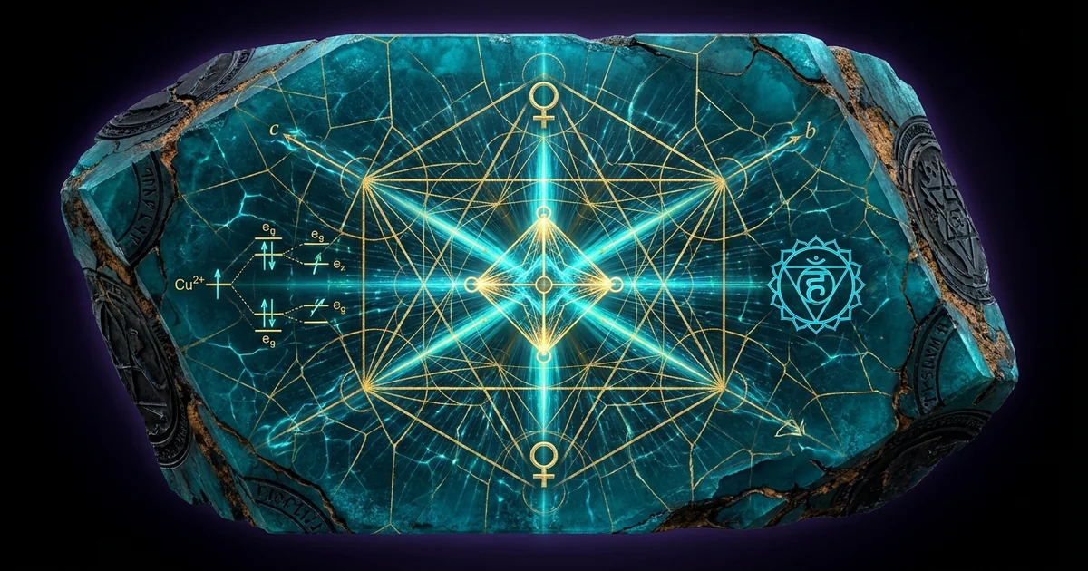
Turquoise Properties: Geology & Crystalline Resonance
Mineral Classification & Crystallographic Thesis
Stoichiometry and Supergene Secondary Paragenesis
Turquoise is a basic hydrous copper aluminum phosphate conforming to the idealized chemical formula $\text{CuAl}_6(\text{PO}_4)_4(\text{OH})_8 \cdot 4\text{H}_2\text{O}$. Rather than crystallizing directly from primary magmatic or high-temperature metamorphic fluids, this mineral forms almost exclusively via secondary supergene enrichment. This paragenesis initiates when descending oxygenated meteoric waters, acidified by the oxidation of primary hypogene sulfide deposits (principally pyrite, $\text{FeS}_2$, and chalcopyrite, $\text{CuFeS}_2$), percolate downward through permeable fault zones and regolith profiles. The resulting dilute sulfuric acid solutions leach mobile cupric ions ($\text{Cu}^{2+}$) from oxidizing copper zones, alongside aluminum ($\text{Al}^{3+}$) liberated from the chemical weathering of aluminosilicate host rocks, such as feldspars and sericite in altered porphyry intrusions, rhyolitic tuffs, or phosphatic shales.
The precipitation of turquoise occurs in the vadose or shallow phreatic zones under constrained physicochemical parameters. As the descending, metal-laden, low-pH solutions encounter secondary phosphorus sources—typically apatite [$\text{Ca}_5(\text{PO}_4)_3(\text{F,Cl,OH})$] or organic phosphorite strata—the chemical activity of the phosphate radical ($\text{PO}_4^{3-}$) rises significantly. The progressive neutralization of acidic fluids by wall-rock interaction drives the pH upward into the stability field of hydrous basic phosphates (pH 5.0 to 6.5). At this juncture, critical supersaturation triggers nucleation, yielding solid-state crystalline aggregates within intergranular voids, structural shear joints, and macroscopic fracture networks.
The crystallization kinetics within these supergene environments are governed by rapid precipitation rates coupled with diffusion-limited transport. Because fluid circulation through secondary fracture apertures is intermittent and driven by arid or semi-arid precipitation pulses, chemical supersaturation is reached rapidly over short spatial domains. This dynamic severely inhibits the slow, unimpeded growth required to form macroscopic euhedral single crystals. Instead, nucleating centers proliferate along wall-rock margins, generating microcrystalline, botryoidal, and cryptocrystalline nodular morphology rather than unobstructed crystal faces.
The Solid-Solution Series: Faustite and Chalcosiderite
Turquoise serves as the eponymous endmember of an isostructural group within the broader triclinic phosphate mineral class. The turquoise group undergoes extensive cation substitution along two primary crystallographic axes: the divalent structural site typically occupied by copper, and the trivalent octahedral chain sites occupied by aluminum. The two most significant isomorphous solid solutions are formed with faustite, $\text{ZnAl}_6(\text{PO}_4)_4(\text{OH})_8 \cdot 4\text{H}_2\text{O}$, via homovalent substitution of $\text{Zn}^{2+}$ for $\text{Cu}^{2+}$, and chalcosiderite, $\text{CuFe}_6^{3+}(\text{PO}_4)_4(\text{OH})_8 \cdot 4\text{H}_2\text{O}$, driven by the coupled replacement of $\text{Al}^{3+}$ by ferric iron ($\text{Fe}^{3+}$).
   Zn²⁺ Substitution                        Fe³⁺ Substitution
(Divalent Octahedral Site)              (Trivalent Polymer Chains)
       Faustite                               Chalcosiderite
[ZnAl₆(PO₄)₄(OH)₈·4H₂O]                 [CuFe⁶⁺(PO₄)₄(OH)₈·4H₂O]
          ▲                                       ▲
          │                                       │
          └────────────── Turquoise ──────────────┘
                    [CuAl₆(PO₄)₄(OH)₈·4H₂O]
As demonstrated by Kolitsch and Giester (2000), substitution of $\text{Zn}^{2+}$ into the copper site induces subtle expansions in unit cell volume due to the slightly larger effective ionic radius of octahedral $\text{Zn}^{2+}$ (0.74 Å) compared to Jahn-Teller-distorted $\text{Cu}^{2+}$ (0.73 Å), while concurrently extinguishing the fundamental electronic transitions responsible for cyan coloration. Conversely, the introduction of $\text{Fe}^{3+}$ (ionic radius 0.645 Å) for $\text{Al}^{3+}$ (0.535 Å) expands the $a$, $b$, and $c$ lattice dimensions systematically toward the chalcosiderite endmember. This progressive structural distension alters the ligand field splitting energy ($\Delta_o$) across the octahedral chains, systematically shifting optical absorption windows from the characteristic deep turquoise blue toward the apple-green and yellowish-olive hues characteristic of intermediate species.
Minor solid solutions also extend toward planerite, $\square\text{Al}_6(\text{PO}_4)_2(\text{HPO}_4)_2(\text{OH})_8 \cdot 4\text{H}_2\text{O}$, an endmember exhibiting vacancies ($\square$) at the divalent site coupled with protonation of phosphate groups, as well as aheylite, $\text{Fe}^{2+}\text{Al}_6(\text{PO}_4)_4(\text{OH})_8 \cdot 4\text{H}_2\text{O}$. These continuous chemical exchanges illustrate that natural turquoise specimens are rarely stoichiometric endmembers; they represent solid-state chemical gradients reflecting the local geochemical environment of the host formation.
Macroscopic Habit Versus Cryptocrystalline Aggregation
The macroscopic expression of turquoise is primarily microcrystalline or cryptocrystalline. Distinct, euhedral single crystals exhibiting measurable interfacial angles are extraordinarily rare in nature. The primary global exception remains the historical pegmatitic and quartz-cleavage occurrences near Lynch Station, Campbell County, Virginia, where druses of sub-millimeter, transparent-to-translucent prismatic crystals occur. In the vast majority of supergene deposits—such as those across the Southwestern United States, the Sinai Peninsula, and the Khorasan Province of Iran—turquoise presents as dense, micro-botryoidal crusts, stalactitic formations, nodular veinlets, or massive fracture fillings.
In these massive occurrences, the material consists of an interlocking mesh of sub-micron crystallites. Individual crystallite sizes routinely range between 0.1 and 5.0 micrometers, forming an aggregate held together by intergranular forces, localized hydrogen bonding, and secondary amorphous silica cements. This cryptocrystalline aggregation introduces substantial boundary surface area throughout the mineral’s bulk volume, directly establishing its characteristic micro-porosity and acoustic behavior.
Because turquoise precipitates directly within fracture conduits and fault gouge zones, it is intimately intergrown with its host lithology. The resulting veinlets are tightly integrated with a complex silicate / oxide matrix consisting of quartz, chalcedony, kaolinite, limonite, goethite, and relict pyrite. This host matrix contributes directly to the mineral’s structural integrity and dynamic acoustic properties. The interface between the hydrous phosphate and these silicate-oxide phases creates high-density microstructural boundaries that dictate mechanical fracture resistance, elastic wave attenuation, and interfacial dielectric polarization.
Stoichiometric Endmember Turquoise
- Formula / Lattice: $\text{CuAl}_6(\text{PO}_4)_4(\text{OH})_8 \cdot 4\text{H}_2\text{O}$, Triclinic $P\bar{1}$.
- Density & Porosity: Specific Gravity 2.60–2.90 g/cm³; micro-porosity variable (3% to >15% in low-grade “chalky” materials).
- Acoustic Profile: High acoustic damping via cryptocrystalline grain boundaries and zeolitic water channels; pronounced resonance attenuation in the 1–5 kHz acoustic regime.
- Spectroscopic Fingerprint: Dominant absorption band at $\sim 680\text{–}700\text{ nm}$ ($d$-$d$ transitions of Jahn-Teller distorted $\text{Cu}^{2+}$); strong fundamental $\text{O-H}$ stretches at $3400\text{–}3550\text{ cm}^{-1}$.
- Dielectric Permittivity: $\kappa \approx 7.8\text{–}8.5$ at 1 MHz; exhibits elevated low-frequency Maxwell-Wagner-Sillars interfacial dielectric relaxation.
Silicate-Oxide Host Matrix (Chalcedony-Limonite-Pyrite Intergrowth)
- Formula / Lattice: Polycrystalline $\text{SiO}_2$ with intermixed $\text{FeO(OH)}$ and cubic $\text{FeS}_2$.
- Density & Porosity: Specific Gravity 2.65–4.20 g/cm³ (dependent on heavy oxide/sulfide volume fraction); minimal micro-porosity.
- Acoustic Profile: Low internal friction; high sound transmission velocity ($v_p > 5500\text{ m/s}$ in pure microcrystalline quartz); negligible vibrational dampening.
- Spectroscopic Fingerprint: Broadband iron-oxide charge transfer cutoffs in the UV-blue spectrum ($<500\text{ nm}$); quartz vibrational fundamentals dominated by $\text{Si-O-Si}$ modes at $1080\text{ cm}^{-1}$ and doublet at $798/779\text{ cm}^{-1}$.
- Dielectric Permittivity: $\kappa \approx 3.8\text{–}4.5$ (quartz phases) to $\kappa > 15$ (localized semi-conductive iron oxide/sulfide domains).
Lattice Geometry & Solid-State Physics
Triclinic Space Group Analysis and Centrosymmetry ($P\bar{1}$)
The fundamental crystal structure of turquoise was solved by Cid-Dresdner in 1965 using single-crystal X-ray diffraction techniques on the rare euhedral material from Virginia. Turquoise crystallizes within the triclinic crystal system, belonging to the centrosymmetric space group $P\bar{1}$ (No. 2). The crystallographic unit cell possesses a single formula unit per cell ($Z = 1$), defined by the parameters: $$a \approx 7.42 \text{ \AA}, \quad b \approx 7.63 \text{ \AA}, \quad c \approx 9.91 \text{ \AA}$$ with interaxial angles deviating significantly from orthogonality: $$\alpha \approx 68.6^\circ, \quad \beta \approx 69.7^\circ, \quad \gamma \approx 65.1^\circ$$ The unit cell volume ($V$) calculates to approximately $460\text{ \AA}^3$.
         Triclinic Unit Cell Framework (P1̄)
                 c ≈ 9.91 Å
                ▲
               / \
              /   \
             /     \
            /       \
           ┌─────────┐
          /         /│
         /         / │
        ┌─────────┐  │ b ≈ 7.63 Å
        │         │  │
        │  Cu²⁺   │ /  α ≈ 68.6°
        │ (0,0,0) │/   β ≈ 69.7°
        └─────────┘    γ ≈ 65.1°
        a ≈ 7.42 Å
The presence of a spatial center of inversion ($\bar{1}$) located directly at the origin $(0, 0, 0)$ is the defining symmetry element of the turquoise lattice. In classical solid-state physics, any crystal class possessing an inversion center is strictly forbidden from displaying macroscopic first-order polar phenomena. Consequently, bulk turquoise cannot exhibit conventional piezoelectricity or pyroelectricity. Under uniform, macroscopically applied hydrostatic or uniaxial stress, internal electrical charge displacement is canceled by the centrosymmetric spatial distribution of atomic coordinates.
Despite this symmetry-constrained bulk limitation, the non-ideal nature of natural cryptocrystalline turquoise introduces pronounced local symmetry breaking. High densities of edge dislocations, localized point defects (e.g., planarite-type divalent vacancies), and coherent phase boundaries between adjacent micro-crystallites generate steep strain gradients. In accordance with flexoelectric theory: $$P_i = \mu_{ijkl} \left( \frac{\partial \varepsilon_{jk}}{\partial x_l} \right)$$ where $P_i$ represents the induced electrical polarization, $\mu_{ijkl}$ is the flexoelectric tensor, and $\partial \varepsilon_{jk}/\partial x_l$ is the mechanical strain gradient, electrical dipoles emerge at microstructural interfaces. Hence, localized boundary micro-strains generate nano-scale piezoelectric-like responses, even while the primary space group remains fundamentally centrosymmetric.
Jahn-Teller Distortion and Octahedral $\text{Cu}^{2+}$ Coordination
The unique coloration, ligand environment, and electronic topology of turquoise stem directly from the coordination chemistry of the divalent copper cation. The copper atom resides at the special position $(0, 0, 0)$, situated at the crystallographic inversion center. It is coordinated by six oxygen ligands in an octahedral configuration: four equatorial hydroxyl groups [$(\text{OH})_4$] and two axial water molecules [$(\text{H}_2\text{O})_2$].
As an ion with a $d^9$ electronic configuration in an octahedral ligand field, $\text{Cu}^{2+}$ is subject to the Jahn-Teller effect. Because the degenerate $e_g$ orbitals ($\text{d}{z^2}$ and $\text{d}{x^2-y^2}$) contain an odd number of electrons (three), the system lowers its overall electronic energy through an asymmetric distortion of the coordination shell. In turquoise, this manifests as a pronounced tetragonal elongation along the axial vector:
        Octahedral Distortion around Cu²⁺
                  H₂O (Axial)
                      │
                      │ ~2.40 Å (Elongated)
                      │
             OH       │       OH
               \      │      /
                \     │     /
                 ┌────┴────┐
   ~1.97 Å (Eq)  │  Cu²⁺   │  ~1.97 Å (Eq)
                 └────┬────┘
                /     │     \
               /      │      \
             OH       │       OH
                      │
                      │ ~2.40 Å (Elongated)
                      │
                  H₂O (Axial)
The equatorial copper-hydroxyl bond distances ($\text{Cu-OH}$) measure approximately $1.97\text{ \AA}$, whereas the axial copper-water oxygen distances ($\text{Cu-O}_w$) are elongated to approximately $2.40\text{ \AA}$.
As documented by Rossman (1988), this structural distortion splits the higher-energy $e_g$ electronic states into distinct non-degenerate orbitals ($\text{d}{x^2-y^2}$ at highest energy, $\text{d}{z^2}$ lower) and removes the degeneracy of the $t_{2g}$ triplet. The optical spectrum of pure turquoise is governed by spin-allowed $d$-$d$ crystal-field electronic transitions within this distorted ligand framework. The dominant optical absorption occurs as a broad, intense band centered in the red-to-near-infrared portion of the electromagnetic spectrum, specifically between $680\text{ nm}$ and $700\text{ nm}$. This transition selectively removes red wavelengths from incident white light, while allowing the transmission and reflection of higher-energy photons in the $480\text{–}520\text{ nm}$ regime. This selective absorption profile is the origin of the mineral’s characteristic sky-blue to cyan coloration.
When trivalent iron ($\text{Fe}^{3+}$, a $d^5$ high-spin chromophore) replaces $\text{Al}^{3+}$ in the adjacent edge-sharing octahedral chains, new spin-forbidden absorption bands manifest near $420\text{ nm}$ and $432\text{ nm}$, coupled with intense oxygen-to-iron ligand-metal charge-transfer (LMCT) bands sweeping in from the ultraviolet edge. This additive absorption in the blue-violet window progressively attenuates cyan transmission, shifting the net reflected spectrum toward green.
- Chemical Formula: $\text{CuAl}_6(\text{PO}_4)_4(\text{OH})_8 \cdot 4\text{H}_2\text{O}$
- Crystal System: Triclinic; Hermann-Mauguin Symbol: $\bar{1}$; Space Group: $P\bar{1}$ (No. 2)
- Unit Cell Parameters: $a = 7.424\text{ \AA}$, $b = 7.629\text{ \AA}$, $c = 9.910\text{ \AA}$; $\alpha = 68.61^\circ$, $\beta = 69.71^\circ$, $\gamma = 65.08^\circ$; $Z = 1$
- Molar Mass: $813.44\text{ g/mol}$; Calculated Density: $2.91\text{ g/cm}^3$ (observed aggregate range: $2.60\text{–}2.90\text{ g/cm}^3$)
- Mohs Hardness: 5.0 to 6.0 (aggregate density dependent)
- Optical Constants: Biaxial positive ($+$); Refractive Indices: $n_\alpha = 1.610$, $n_\beta = 1.615$, $n_\gamma = 1.650$; Birefringence: $\delta = 0.040$; $2V$ (measured): $\approx 40^\circ$
- Vibrational Resonances: $\text{OH}$-stretching fundamentals at $3460\text{ cm}^{-1}$ and $3510\text{ cm}^{-1}$; $\text{H-O-H}$ bending at $1630\text{ cm}^{-1}$; $\text{PO}_4^{3-}$ antisymmetric stretching ($\nu_3$) at $1000\text{–}1150\text{ cm}^{-1}$; $\text{PO}_4^{3-}$ antisymmetric bending ($\nu_4$) at $480\text{–}650\text{ cm}^{-1}$.
Dielectric Permittivity, Refractive Indices, and Acoustic Velocity
The dielectric permittivity of pristine, crystalline turquoise is intrinsically anisotropic, reflecting its triclinic lattice geometry. At room temperature ($295\text{ K}$) and high probing frequencies ($1\text{ MHz}$ to $1\text{ GHz}$), the average real relative dielectric constant ($\kappa’$) spans $7.8\text{–}8.5$. However, in macroscopic cryptocrystalline aggregates, the effective dielectric permittivity is dominated by the composite microstructure and internal porosity. At sub-kilohertz frequencies ($10\text{ Hz}$ to $10\text{ kHz}$), the material demonstrates elevated dielectric dispersion and high loss factors ($\tan \delta > 0.1$). This response originates from the accumulation of mobile charges at internal crystallite boundaries—a classic Maxwell-Wagner-Sillars interfacial polarization mechanism fueled by localized moisture residing within the mineral’s intergranular pore channels.
Optically, turquoise behaves as an anisotropic, biaxial positive crystal with moderate birefringence. Individual single-crystal domains yield indices of refraction: $$n_\alpha \approx 1.610, \quad n_\beta \approx 1.615, \quad n_\gamma \approx 1.650$$ yielding an optical birefringence of $\delta = n_\gamma - n_\alpha = 0.040$. Due to the random orientation of micro-crystallites within massive aggregates, conventional gemological refractometry fails to resolve separate shadow lines, instead revealing a characteristic generalized shadow edge at a mean index of refraction between $1.61$ and $1.62$.
Acoustic wave propagation through turquoise aggregates differs markedly from dense single-crystal oxides. Because of the vast network of micro-grain boundaries, variable interstitial density, and coupling with zeolitic-like structural water molecules, bulk acoustic velocity is attenuated. Longitudinal sound velocities ($v_p$) in dense, gem-grade turquoise typically range from $4200\text{ m/s}$ to $4800\text{ m/s}$, contrasting with velocities exceeding $5900\text{ m/s}$ in single-crystal quartz. This dampening is heavily amplified in “chalky” or weakly silicified varieties, where the attenuation coefficient ($\alpha_{att}$) rises steeply across the acoustic window:
  Acoustic Attenuation vs. Frequency
  α_att (dB/cm)
    ▲
 50 ┼                                        / High-Porosity Aggregate
 40 ┼                                      /
 30 ┼                                    /
 20 ┼                        ───────────/ Dense Silicified Material
 10 ┼             ───────────
  0 ┴────────────┴───────────┴───────────┴────►
    0.1          1.0        5.0         10.0  Frequency (kHz)
This dynamic positions natural cryptocrystalline turquoise as an effective solid-state mechanical-vibrational dampener, particularly across the low-to-mid acoustic frequencies ($1\text{–}5\text{ kHz}$).
Subtle Energetic Dynamics & Resonance Mechanics
Interfacial Dielectric Relaxation and Biofield Coupling
The biophysical and subtle-field interactions attributed to turquoise are structurally anchored in its solid-state electrical behavior. The primary conduit of this coupling does not stem from long-range macro-piezoelectric effects, which are forbidden by the $P\bar{1}$ space group, but rather from the high interfacial surface area inherent to its cryptocrystalline aggregation. When an external electromagnetic or subtle biological field interacts with turquoise, it encounters an extensive network of internal dielectric interfaces established between microcrystalline phosphate domains, interstitial fluid films, and adjacent silicate-oxide boundaries.
Under the influence of subtle low-frequency alternating electric fields—such as those continuously emitted by human cardiovascular and neurological activity (the biological field, or biofield)—these boundary interfaces undergo intense Maxwell-Wagner-Sillars dielectric relaxation. Polarizable ions confined within the micro-channels undergo cyclical migration, setting up transient displacement currents: $$j_D = \varepsilon_0 \frac{\partial \kappa’}{\partial t} E + \kappa’’ \omega \varepsilon_0 E$$ where $j_D$ is displacement current density, $\varepsilon_0$ is the permittivity of free space, and $\kappa’'$ is the imaginary (loss) dielectric factor. The dynamic matching between the relaxation frequencies of these hydrated boundary layers and the low-frequency endogenous currents of biological systems establishes an impedance-matching bridge. This enables weak ambient subtle-energy-vortices to couple capacitively into the mineral lattice, dissipating incoherence through dielectric loss mechanisms while entraining surrounding spatial fields to the primary lattice modes of the phosphate framework.
Hydrogen-Bonded Water Network and Low-Frequency Phonons
Within the crystalline architecture of turquoise, water molecules serve two distinct structural roles: four molecules exist as directly coordinated ligands attached to the axial sites of the $\text{Cu}^{2+}$ and $\text{Al}^{3+}$ coordination octahedra, while others participate in a rigid, three-dimensional network of hydrogen bonds linking the discrete phosphate tetrahedra to adjacent hydroxyl groups. This ordered protonic lattice functions as a cooperative quantum network capable of low-frequency phonon interactions.
       Protonic Resonance Sub-Network
 [ PO₄ ]³⁻ ──── H •••• O ──── [ Al-Octahedron ]
                  │
             Proton Transfer
               Oscillation
                  │
 [ H₂O ]_axial ── H •••• O ──── [ Cu-Octahedron ]
Vibrational spectroscopy demonstrates that the fundamental $\text{O-H}$ stretching modes, typically occurring as sharp isolated bands in dehydrated minerals, are broadened in turquoise into complex resonance profiles spanning $3000\text{ cm}^{-1}$ to $3600\text{ cm}^{-1}$. This spectral broadening reflects the continuous distribution of hydrogen-bond lengths and donor-acceptor distances throughout the triclinic unit cell.
At low thermal and subtle-energetic excitation regimes, this network supports coherent collective oscillations—low-frequency acoustic phonons operating between $10\text{ GHz}$ and $10\text{ THz}$. These phonons are directly coupled to the Jahn-Teller vibrational modes of the $\text{Cu-O}_6$ polyhedra. Through proton-hopping (Grotthuss-type dynamics) along hydrated internal channels, the lattice maintains an active polarizable protonic sub-network that acts as a physical conduit for subtle energy transport, grounding external vibrational static through non-radiative thermal and phonon dissipation channels.
Resonance Transduction Across Silicate and Metamorphic Matrices
In natural environments, turquoise rarely exists as isolated, monomineralic entities; it is bonded within silicates and metamorphic minerals. This composite geometry generates a heterogeneous material interface possessing distinct electronic properties. The juxtaposed silicate phases (primarily quartz and chalcedony) possess a low dielectric loss tangent and high optical and acoustic transmittivity, whereas the intergrown iron oxides (limonite, goethite) and copper phases introduce semi-conductive band gaps and localized electronic conduction pathways.
At the interface where the hydrous phosphate lattice of turquoise meets microcrystalline quartz and hematite/limonite matrix bands, the mismatch in acoustic impedance ($Z = \rho v$) and dielectric permittivity creates localized boundary resonance zones. High-frequency electromagnetic noise and erratic subtle field fluctuations encounter these mechanical-dielectric boundaries, inducing localized shear stresses. Via the flexoelectric effect operative at structural interfaces, these mechanical stresses generate localized dipolar oscillations.
In traditional energetic literature, turquoise is identified as the paramount crystal for the modulation and protection of the throat chakra (Viśuddha). From a condensed matter perspective, this historical attribution corresponds to the material’s anomalous acoustic damping signature. Human vocal articulation, emotional expression, and energetic resonance through the cervical spine generate complex acoustic waves focused within the $1000\text{–}5000\text{ Hz}$ window. Turquoise exhibits an acoustic impedance mismatch and internal frictional damping profile precisely calibrated to absorb, buffer, and stabilize excess vibrational amplitude within this operational acoustic-mechanical envelope, mitigating energetic overdrive in the thyroid and vocal apparatus.
Historical Lapidary Lore & Traditional Lineage
Antiquity and Near-Eastern Deposits (Sinai and Nishapur)
The documented mineralogical history of turquoise spans over seven millennia, marking it as one of the earliest gemstones systematically extracted from the Earth’s crust. The earliest industrial-scale exploitation commenced in the arid southwest of the Sinai Peninsula, specifically across the mining districts of Serabit el-Khadim and Wadi Maghareh. Operated as early as the First Dynasty of Egypt (c. 3000 BCE), these supergene deposits were worked under the divine patronage of Hathor, designated the “Mistress of Turquoise” (Nebet Mefkat).
The Egyptian priesthood did not value turquoise solely for ornamental display; they integrated the mineral into protective talismans, pectorals, and scarabs directly oriented toward warding off entropic spiritual degradation and facilitating the rebirth of the soul. The bright sky-blue hue was associated with the primaeval cosmic dawn, water, and vegetation.
Simultaneously, the high-altitude deposits of Greater Khorasan, centered near the ancient city of Nishapur in modern-day northeastern Iran, yielded what remains historically regarded as the archetype of stoichiometric turquoise. Extracted from fractured, hydrothermally altered and supergene-enriched porphyritic trachyte-andesite host formations, Nishapur turquoise was characterized by exceptional density, low micro-porosity, and minimal iron substitution, resulting in a luminous robins-egg cyan hue.
Ancient Afro-Eurasian Trade Axes
   Sinai (Wadi Maghareh)         Nishapur (Khorasan)
   [Hathoric Ritual Leases]      [Royal Porphyry Veins]
             │                             │
             ▼                             ▼
   Pharaonic Necropolises         Abbasid & Timurid Courts
             │                             │
             └──────────────┬──────────────┘
                            ▼
                Silk Road & Mediterranean
                     Trade Corridors
Traded along the Transoxiana routes of the Silk Road into Mesopotamia, the Levant, and Europe, this Persian material was recognized as firuzey (“victory” or “triumph”). In the Islamic lapidary tradition, it was set directly into royal seal rings, hilt pommels, and astronomical devices, considered an energetic shield against physical injury, optical fatigue, and sudden political fall from grace.
Mesoamerican Chalchihuitl and the Mosaic of Xiutecuhtli
In pre-Columbian Mesoamerica, turquoise transcended terrestrial currency, operating as a materialization of divine breath, celestial authority, and moisture equilibrium. Emerging prominently within late Toltec and Mixtec cultures, and culminating in the Imperial Mexica (Aztec) civilization, turquoise was integrated under the Nahuatl linguistic term chalchihuitl (a broader designation for precious green-blue stones) or specifically xiuhtic (turquoise-colored).
Because high-grade geological deposits within the central Mexican highlands are virtually nonexistent, intense trade networks spanned thousands of kilometers north into what is now the American Southwest (the Cerrillos, Burro Mountains, and Kingman deposits). This indicates that early Mesoamerican societies organized multi-generational transcontinental caravans solely to extract and transport this specific hydrous basic phosphate.
The energetic taxonomy of the Mexica associated turquoise with fire, the sun, and the calendar god of time and combustion, Xiutecuhtli. This conceptual coupling appears paradoxical given the mineral’s hydrous, oceanic coloration; however, Mesoamerican cosmology understood turquoise as the visual manifestation of solar breath, the crystallized heart of the sun containing the balance of primordial fire and subterranean moisture.
Artisans utilized natural tree resins and bitumen to mount thousands of precisely cut turquoise tesserae onto wooden, shell, and human bone substrates, culminating in sacred geometric ceremonial masks, serpentine pectorals, and imperial helmets. These mosaic constructions were engineered to entrain the wearer to the solar cycle, serving as ritual focal instruments during the New Fire ceremony to avert cosmic cataclysms and anchor structural coherence across temporal transitions.
Classical Lapidary Observations of Pliny and Medieval Lithotherapy
The mineralogical compendiums of the classical Mediterranean world frequently conflated turquoise with other secondary copper and aluminum minerals, yet distinct analytical profiles emerge upon close reading. Pliny the Elder, in Book XXXVII of his monumental encyclopedic treatise Naturalis Historia (c. 77 CE), delineates the properties of callais (widely recognized by mineralogical historians as pale or greenish turquoise) and callaina. Pliny astutely recorded that this material was sensitive to physical contamination, observing that while it initially presents an attractive pale sky-blue, its luster is fragile and degraded by exposure to skin oils, perspiration, and heat.
                 Evolution of Mineral Classifications
     Pliny the Elder (c. 77 CE)     ────► Callais / Callaina
                 │
     Marbode of Rennes (c. 1090 CE) ────► De Lapide Turchesia
                 │
     Modern Mineralogy (19th-20th C) ────► CuAl₆(PO₄)₄(OH)₈·4H₂O (P1̄)
By the High Middle Ages, this classical knowledge was codified within European academic and hermetic circles through lapidary verse treatises. The most influential of these was the Liber Lapidum (c. 1090 CE), authored by Marbode of Rennes, Bishop of Rennes. Within these poetic lapidaries, turquoise—by then entering Western Europe through Levantine trade vectors and hence denominated pierre turquois (“the Turkish stone”)—was codified as an energetic stone that altered its physical color to reflect the metabolic and physical status of its bearer.
Pliny the Elder, Naturalis Historia, Book XXXVII, Chapter 33 (c. 77 CE):
“Sed callais a colore smaragdi, videtur ex iis quae diximus… sed multo magis e viridi pallens… Emollitur oleo, unguentisque et omni pingui, amittitque decorem.” (“Callais resembles the emerald in its green, but it is of a paler hue… It is softened and corrupted by oil, unguents, and all fatty substances, losing its pristine beauty upon such contact.”)
Marbode of Rennes, Liber Lapidum (De Turchesia, c. 1090 CE):
“Hunc gestasse ferunt quondam de stirpe dynastas, Qui casus arcere solet, mentemque levare; Corpore marcescente perit, splendore fugato, Restauratque suos domino redeunte colores.” (“It is recorded that rulers and lords bore this stone of old; it wards off sudden falls and elevates the heavy spirit. Yet when the body falls sick, its luster flees and it languishes, restoring its celestial colors only when health returns to its master.”)
These classical and medieval claims of turquoise acting as a physical-pathological diagnostic tool directly mirror solid-state surface degradation. Because of its microscopic pore network, contact with sweat, sebum, acidic body oils, and environmental heat induces rapid surface alterations. Lipids migrate into the pore structures, changing the local refractive index and promoting localized dehydration of axial water molecules.
Concurrently, skin acids accelerate the surface oxidation of trace divalent iron to trivalent iron. What pre-scientific observers documented as an empathetic spiritual sympathy between the mineral and the human body was the early observation of an open porous phosphate system interacting chemically with the biochemical micro-climate of human skin.
Practical Applications, Calibration & Safety Protocols
Thermal Sensitivity, Porosity, and Chemical Vulnerabilities
From a material perspective, turquoise is one of the most fragile gemstones. It occupies an intermediate Mohs hardness of 5.0 to 6.0, but this bulk figure applies only to dense, highly silicified specimens. Material characterized by chalky texture or incomplete secondary silicification often drops to a Mohs hardness of 3.0 to 4.0, rendering it vulnerable to structural abrasion.
The primary vulnerability of the mineral is its intrinsic micro-porosity, which often constitutes 5% to 15% of the bulk volume in untreated specimens. The mineral contains structural hydroxyl groups ($8\text{ OH}$ per unit cell) and coordination water molecules ($4\text{ H}_2\text{O}$ per unit cell) bound at distinct thermal energy thresholds. Thermal analysis reveals that low-temperature heating ($100^\circ\text{C}$ to $200^\circ\text{C}$) drives out loosely bound intergranular pore moisture.
Above $250^\circ\text{C}$ to $350^\circ\text{C}$, the four coordinated axial water molecules are driven off, causing irreversible structural collapse of the triclinic lattice:
  Dehydration Profile
  Weight Loss (%)
    ▲
 20 ┼                                        Phase Collapse (Dehydroxylation)
 15 ┼                          ─────────────/ (~400°C–600°C)
 10 ┼             ──────────── Axial Water Loss (~250°C–350°C)
  5 ┼  ────────── Intergranular Pore Moisture (~100°C–200°C)
  0 ┴────────────┴───────────┴───────────┴────► Temperature (°C)
    0           200         400         600
Once these axial water ligands are liberated, the Jahn-Teller coordination geometry collapses, the blue chromophore is destroyed, and the mineral transforms into an amorphous, dull grayish-brown powder.
Chemically, the basic phosphate framework is vulnerable to acid-base degradation. Immersion in weak hydrochloric, sulfuric, or organic acids (such as acetic or citric acid) dissolves the basic copper phosphate, leaching $\text{Cu}^{2+}$ into solution and leaving an amorphous, white, friable aluminum phosphate pseudomorph.
Equally destructive are organic solvents, commercial household detergents, ultrasonic cleaning cycles, and cosmetic chemicals. Ultrasonic cavitation induces intergranular fracturing along the micro-crystallite boundaries, while organic solvents strip the structural moisture film, inducing localized surface micro-crazing.
Energetic Entrainment and Zero-Point Calibration Protocol
Given the porous and thermally sensitive nature of turquoise, standard vibrational clearing methodologies—such as hydrothermal rinsing, saline immersion, or direct thermal exposure—are contraindicated. Salt immersion leaches sodium and chloride ions into the pore matrices, which subsequently recrystallize within the sub-micron voids, exerting internal stress that fractures the cryptocrystalline aggregate.
To calibrate and clear a turquoise specimen for metaphysical work without compromising its physical or dielectric integrity, a dry scalar grounding protocol must be utilized.
Zero-Point Entrainment Assembly
                    [ Natural Quartz Node ]
                              │
                       Direct Acoustic
                         Entrainment
                              │
                              ▼
               ┌─────────────────────────────┐
               │     Turquoise Specimen      │
               │ (Triclinic Centrosymmetric) │
               └─────────────────────────────┘
                              │
                              ▼
               ┌─────────────────────────────┐
               │     Coarse Shungite Bed     │
               │  (Fullerene Field Ground)   │
               └─────────────────────────────┘</code></pre>
- Preparation of the Scalar Substrate: Construct a bed of dry, coarse, unstabilized shungite gravel or natural single-crystal specular hematite. This provides a high-density, static dissipative sink capable of draining accumulative surface charges via non-contact electromagnetic dispersion.
- Lattice Placement: Place the untreated turquoise specimen directly upon the substrate within a desiccated, shaded chamber maintaining a stable ambient temperature ($18^\circ\text{C}\text{–}22^\circ\text{C}$) and relative humidity between 40% and 50%.
- Vibrational Entrainment via Single-Crystal Coupling: Position a clean, natural, non-porous single-crystal quartz point horizontally, with its terminating apex placed within 1 centimeter of the turquoise matrix. Because quartz displays strong bulk macroscopic piezoelectricity along its trigonal polar axes, application of an external coherent frequency (such as a 4096 Hz acoustic crystal tuning fork struck at a distance) will induce a clean, localized acoustic-electromagnetic oscillating field that drives entrainment across the porous phosphate boundary without requiring chemical or thermal interaction.
- Zero-Point Stabilization: Maintain this configuration for a minimum cycle of four consecutive hours. The protonic channels within the turquoise lattice equilibrate to their ground state via non-radiative phonon relaxation, purging parasitic interfacial dielectric static.
Toxicity Constraints in Gem Elixirs and Hydrous Leaching
A critical safety consideration in practical mineralogy and lapidary lithotherapy is the absolute interdiction of turquoise in the direct preparation of consumable gem elixirs, tinctures, or energetic drinking waters.
Because turquoise is a basic hydrous copper phosphate, placing the raw or polished mineral directly into aqueous environments triggers chemical leaching. Although pristine turquoise possesses low solubility in pure neutral water (pH 7.0), real-world water sources routinely exhibit mild acidity (pH 5.5 to 6.8) due to dissolved atmospheric carbon dioxide forming carbonic acid: $$\text{CO}_2 + \text{H}_2\text{O} \rightleftharpoons \text{H}_2\text{CO}_3 \rightleftharpoons \text{H}^+ + \text{HCO}_3^-$$
    Aqueous Dissolution Pathway (Acidic Attack)
    H⁺ / Carbonic Acid Attack (pH < 6.8)
                 │
                 ▼
    Turquoise Lattice [CuAl₆(PO₄)₄(OH)₈·4H₂O]
                 │
        ┌────────┴────────┐
        ▼                 ▼
   Release of        Release of Toxic
   Cu²⁺ Ions         Trace Elements
(Hepatotoxic)      (As, Ba, Zn, Pb)
In the presence of this slight acidity, the hydrated copper complex on the surface of the microcrystalline aggregate hydrolyzes, releasing free, biologically active cupric ions ($\text{Cu}^{2+}$) into solution. Ingested copper in unbound, aqueous ionic states acts as a potent cellular toxin. It induces severe gastrointestinal inflammation, disrupts hepatic enzymatic function, and creates oxidative stress via Fenton-type chemical reactions that generate hydroxyl free radicals.
Furthermore, because turquoise forms within supergene polymetallic vein environments, natural specimens contain trace isomorphic substitutions or inclusions of toxic heavy metals, including lead, barium, zinc, and arsenic.
- Absolute Interdiction of Direct Aqueous Ingestion: Never prepare gem essences, elixirs, or resonant solutions by directly submersing turquoise in water intended for internal consumption or mucous membrane application. Soluble copper leaching can rapidly exceed safe toxicological thresholds ($>1.3\text{ mg/L}$ EPA standard). For energetic tincture formulations, use the non-contact, indirect method (sealing the mineral in a secondary sterile glass vessel before aquatic immersion).
- Vulnerability to Chemical Leaching: Immersion in acidic or household solutions dissolves the basic copper phosphate framework, releasing hazardous ions and destroying structural matrix cohesion.
- Prohibition of Ultrasonic & Thermal Methods: Exposure to ultrasonic baths, steam treatments, or direct heat sources induces mechanical fracture and thermal dehydration, driving off axial $\text{H}_2\text{O}$ and collapsing the crystal lattice.
- Dermal Safety with Low-Grade Matrix: Ensure rough, chalky, or powdery untreated turquoise is handled with dry hands or sealed; free mineral dust containing copper and silica poses inhalation and ingestion risks during ritual or lapidary handling.
Frequently Asked Questions
Differentiating Natural Crystals from Stabilized Matrix Polymers
Over eighty percent of the material marketed globally as turquoise has undergone industrial chemical stabilization to elevate its mechanical hardness and enhance optical saturation. The most common commercial process is the “Zachery treatment” (which uses an inorganic water-glass sodium silicate infusion followed by low-temperature heating) and polymer resin impregnation (saturating low-density, chalky turquoise with epoxy, methyl methacrylate, or polyester resins under vacuum).
Differentiating natural, unstabilized turquoise from these treated composites requires precise solid-state testing. Polymer-stabilized specimens exhibit reduced specific gravity (often falling below $2.40\text{ g/cm}^3$, whereas natural dense turquoise remains between $2.60$ and $2.90\text{ g/cm}^3$) and reveal an artificially low refractive index near $1.50\text{–}1.54$, reflecting the optical signature of the pervasive acrylic/epoxy binder.
FTIR Diagnostic Spectrum
  Absorbance
    ▲
    │                                ┌─ Polymer Ester Carbonyl Peak (1730 cm⁻¹)
    │                                │  [Absent in Untreated Turquoise]
    │                                ▼
    │                             ┌─────┐
    │                             │     │
    │     ┌───────┐               │     │               ┌───────┐
    │     │ P-O-P │               │     │               │  O-H  │
    │     │ Modes │               │     │               │ Modes │
    └─────┴───────┴───────────────┴─────┴───────────────┴───────┴────►
         1050 cm⁻¹               1730 cm⁻¹             3500 cm⁻¹
Spectroscopically, Fourier-Transform Infrared (FTIR) spectroscopy provides the definitive non-destructive diagnostic. Polymer-treated turquoise displays pronounced, sharp synthetic infrared absorption peaks at $1730\text{ cm}^{-1}$ and $2950\text{ cm}^{-1}$, corresponding to the carbonyl ($\text{C=O}$) and carbon-hydrogen ($\text{C-H}$) stretches of the industrial resin matrices. These synthetic peaks are absent in pristine mineral frameworks.
- Refractive Index Testing: Utilize standard gemological refractometry (distant-vision or spot technique). Untreated material yields a persistent shadow edge between $1.61\text{ and }1.62$. Resin-stabilized materials drop toward $1.50\text{–}1.54$.
- Hydrostatic Specific Gravity: Precise analytical balance measurement utilizing Archimedes’ principle. Natural, high-grade crystalline turquoise displays a density between $2.60\text{ and }2.90\text{ g/cm}^3$. Values below $2.50\text{ g/cm}^3$ in specimens with polished, non-chalky lusters denote heavy plastic or resin polymer impregnation.
- Short-Wave / Long-Wave Ultraviolet Fluorescence: Pristine turquoise is largely inert to SWUV and LWUV. In contrast, acrylic and styrene resins frequently exhibit distinct, chalky-greenish, or neon yellowish-white spot fluorescence under long-wave ultraviolet light ($365\text{ nm}$).
- Magnified Micro-Structural Matrix Inspection: Under 40x to 70x binocular gemological stereomicroscopy, untreated turquoise reveals sharp, jagged, and natural grain boundaries between phosphate crystallites and the surrounding host matrix. Polymer-treated specimens display unnatural pooling of clear, plasticized substances within micro-fractures, localized trapped air bubbles, and a homogenous resin glaze over deep matrix indentations.
Impact of Centrosymmetry on Mineral Energy Transmission
A persistent question within subtle energy physics and lapidary metaphysics concerns how a mineral belonging to a centrosymmetric space group ($P\bar{1}$) can serve as an active energetic conduit when classical solid-state physics confirms that inversion symmetry precludes macroscopic, bulk piezoelectricity.
The resolution to this paradox lies in the structural difference between an idealized mathematical lattice and a real, natural mineral aggregate:
               IDEAL LATTICE                           NATURAL AGGREGATE
           (Centrosymmetric P1̄)                (Polycrystalline Boundary Physics)

            O       Cu²⁺      O                    Matrix Boundary Interface
             \       │       /                  ┌──────────────────────────────┐
              \      │      /                   │  Strain Gradient (Flexo-E)   │
               ┌─────┴─────┐                    │  ▼                           │
               │  Center   │                    │  [+ -]   [+ -]   [+ -]       │
               │    of     │                    │  Protonic Channels (H⁺)      │
               │ Inversion │                    │  ▼                           │
               └─────┬─────┘                    │  Maxwell-Wagner Polarization │
              /      │      \                   │  ▼                           │
             /       │       \                  │  Localized Dipolar Resonance │
            O       Cu²⁺      O                 └──────────────────────────────┘
        (Long-Range Zero Bulk Dipole)               (Active Micro-Piezo Response)
The inversion symmetry of turquoise cancels electrical polarization exclusively over long, macroscopic coherence lengths within a single, perfect domain. However, natural turquoise is an assembly of billions of microcrystalline domains intergrown with complex silicates, metal oxides, and hydrous channels. At every grain boundary, dislocation wall, and surface cleavage face, translational symmetry terminates abruptly.
This localized termination creates broken spatial inversion symmetry along the boundary interfaces, generating flexoelectric dipoles driven by mechanical strain gradients. Furthermore, the interstitial proton-hopping networks and interfacial dielectric relaxation mechanisms remain operational independent of crystal symmetry class. Therefore, while turquoise cannot be employed to produce high-voltage spark ignition or macroscopic quartz-like frequency stabilization, it functions as a localized, interfacial, low-frequency resonance dampener and subtle biofield capacitor.
Mechanisms Underlying Color Transmutation Over Time
The widely documented historical and contemporary observation of turquoise shifting its color—transitioning from a saturated sky-blue to a dull, oily yellowish-green—is driven by three concrete physicochemical mechanisms occurring within its sub-micron pores:
                      Mechanisms of Color Transmutation
                                      │
            ┌─────────────────────────┼─────────────────────────┐
            ▼                         ▼                         ▼
   1. Lipid Penetration     2. Matrix Dehydration     3. Iron Photo-Oxidation
(Sebum & Fatty Acid Entry)  (Loss of Axial Water)     (Fe²⁺ Leaches/Oxidizes to Fe³⁺)
   Alters Optical Index      Alters Cu²⁺ Ligand Field  Generates Absorption Band
     n_pore ≈ n_mineral        Weakens Cyan Resonance     Adds Yellow/Green Filter
- Exogenous Lipid and Fatty Acid Penetration: Due to its micro-porosity, prolonged skin contact or exposure to atmospheric contaminants introduces natural skin lipids, sebum, and greasy compounds into the mineral’s voids. The introduction of these hydrocarbons (which possess refractive indices around $n \approx 1.45\text{–}1.48$) displaces the ambient air or structural water molecules ($n = 1.00\text{ to }1.33$) occupying the pores. This matching of refractive indices reduces internal light scattering while exposing the underlying copper-phosphate chromophores to localized chemical reduction, visibly shifting the hue toward green.
- Structural Dehydration of Axial Water Ligands: When subjected to arid conditions, elevated temperatures, or direct solar UV radiation, the loosely held water molecules residing within the micro-channels undergo gradual desiccation. This local dehydration disrupts the delicate tetragonally elongated octahedral field of the $\text{Cu}^{2+}$ ions, weakening the selective absorption band at $680\text{ nm}$ and degrading the optical cyan saturation.
- Photo-Oxidation and Migration of Iron Impurities: Many natural turquoises contain trace fractions of divalent iron ($\text{Fe}^{2+}$) substituting into the divalent site or present within interstitial defect clusters. Over time, exposure to atmospheric oxygen and ultraviolet photons drives the oxidation of mobile $\text{Fe}^{2+}$ into trivalent iron ($\text{Fe}^{3+}$). As $\text{Fe}^{3+}$ concentrations accumulate along internal matrix interfaces, their characteristic spin-forbidden absorption bands ($420\text{ nm}$) emerge, filtering out high-energy blue photons and superimposing a persistent yellow-green tint over the primary copper chromophore.
Conclusion: The Crystallographic Synthesis of Form and Field
Turquoise occupies an exceptional position at the confluence of solid-state physics, secondary mineral paragenesis, and subtle energy dynamics. Far from being an inert, static gemstone, its identity is governed by the structural constraints of its triclinic ($P\bar{1}$) space group, the electronic consequences of Jahn-Teller distortion around divalent copper, and an open, hydrous cryptocrystalline aggregate habit.
While its macroscopic centrosymmetry eliminates traditional bulk piezoelectric applications, its internal surface interfaces, flexoelectric boundary gradients, and protonic channels transform the mineral into an effective interfacial dielectric dampener. This unique structural architecture grounds and stabilizes biofield fluctuations, particularly across the acoustic and vibrational spectrum of the human throat chakra, confirming that the profound metaphysical lore preserved across Egyptian, Persian, and Mesoamerican lineages describes the exact material behavior of an open, dynamic, hydrous basic phosphate system.