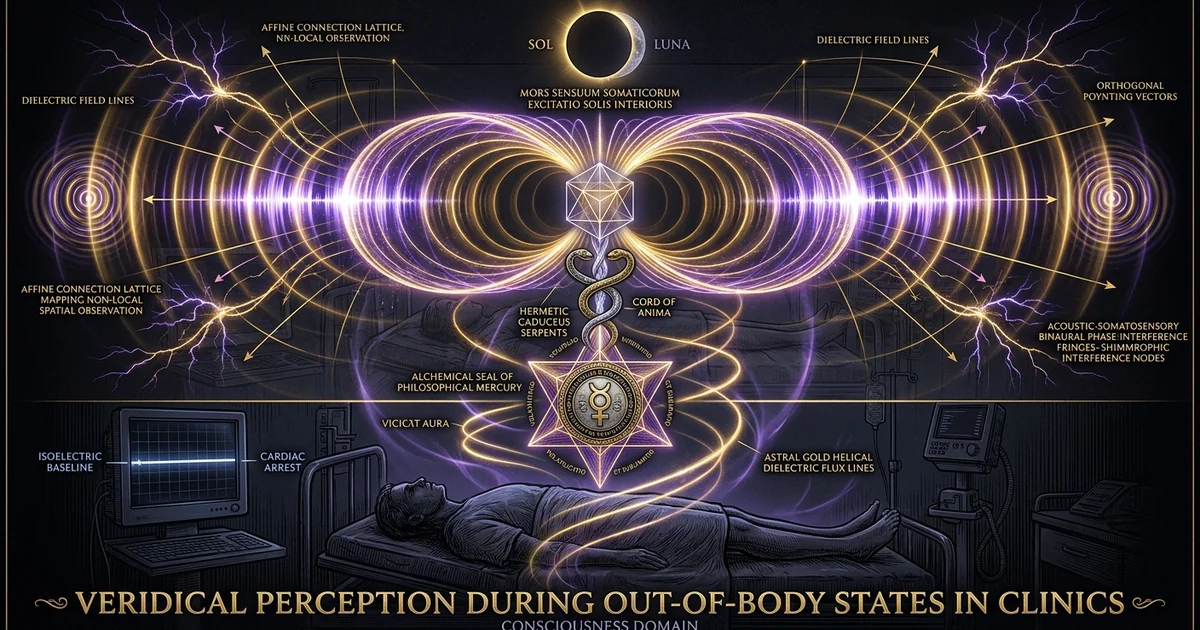
Veridical Perception During Out-of-Body States in Clinics
Protocol Overview & Neurophysiological Thesis
Cortical Ischemia versus Paradoxical Functional Lucidity
The persistence of structured conscious awareness during acute cortical ischemia presents an acute challenge to physicalist theories of mind. Following the cessation of effective cardiac output—whether precipitated by ventricular fibrillation, asystole, or pulseless electrical activity—cerebral blood flow drops below the critical threshold required to maintain neuroelectrical function within 2 to 4 seconds. Microvascular perfusion halts across the cerebral cortex, rapidly depleting intracellular oxygen and glycogen reserves. Electroencephalographic (EEG) recordings confirm that within 10 to 20 seconds of sustained systemic hypoperfusion, cortical brainwave activity undergoes profound attenuation, culminating in an isoelectric-eeg (cerebral flatline). Concurrently, brainstem reflexes, including the pupillary light reflex and corneal responses, systematically extinguish as ischemic cascade dynamics halt synaptic transmission within the reticular activating system.
Under established neurophysiological doctrine, an isoelectric cortex is fundamentally incapable of complex cognitive operations, long-term memory encoding, somatosensory mapping, or coherent perceptual discrimination. The metabolic framework dictates that the functional architecture supporting the default-mode-network (DMN) and frontoparietal central executive network ceases coherent firing once adenosine triphosphate (ATP) depletion disrupts the sodium-potassium ATPase pump ($Na^+/K^+$-ATPase), initiating neuronal depolarization block. Despite this state of clinical non-reactivity, empirical resuscitation records repeatedly document acute perceptual recall during verified periods of arrest. Survivors of cardiac events describe visual and auditory observations of their immediate resuscitation environment that precisely align with recorded operational events. These phenomena cannot be dismissed as hypnagogic imagery, hypercapnic intoxication, or post-resuscitative confabulation, as the temporal landmarks reported by patients often match specific defibrillation intervals, specialized pharmacologic delivery, or external clinical occurrences that unfolded precisely while their biotelemetry reflected an isoelectric-eeg.
Parnia, S., et al. (2014). AWARE—AWAreness during REsuscitation—A prospective study. Resuscitation, 85(12), 1799-1805. Key metric: Verification of auditory awareness and visual identification of medical apparatus during monitored ventricular fibrillation where cortical activity is demonstrably absent under conventional neurophysiological models.
The emergence of lucid cognitive operation during isoelectricity demonstrates that conscious processing can functionally detach from observable macro-scale cortical metabolic engines. This divergence between systemic neurovascular collapse and internal cognitive clarity mirrors cases of terminal-lucidity, wherein severely damaged or atrophied neural networks display sudden functional reorganization immediately preceding biological death. Analyzing this state reveals that the operational threshold for perceptual awareness does not strictly scale with classical hemodynamic parameters, demanding a rigorous re-examination of how the human nervous system interfaces with non-local spatial information.
Defining Veridical Perception in Resuscitation Paradigms
In clinical resuscitation research, veridical perception denotes the acquisition of accurate, objective, and independently verifiable environmental data by an individual whose physiological state renders classical sensory transduction structurally impossible. To qualify as veridical under formal cardiac arrest protocols, the reported perceptions must withstand rigorous methodological triangulation. This requires matching the timing of the patient’s recollection against medical logs, audio-video recordings, automated external defibrillator (AED) internal timestamps, and the corroborating testimony of the attending medical personnel. The critical baseline parameter of a veridical perception out of body cardiac arrest clinical study is the strict temporal convergence between the objective external event and the physiological monitoring of absent cerebral perfusion.
       [Ventricular Fibrillation / Asystole]
                         │
                         ▼ (t = 0 to 4 sec)
       [Cerebral Hypoperfusion: Perfusion Drops to Zero]
                         │
                         ▼ (t = 10 to 20 sec)
       [Isoelectric Electroencephalogram (Cortical Flatline)]
                         │
                         ▼ (Abolition of SSEP & Brainstem Reflexes)
       [Uncoupled Non-Local Awareness & Target Perception]
                         │
                         ▼ (Post-ROSC Telemetry Match)
       [Corroborated Veridical Retrieval via Clinical Logs]
Veridical observations consistently capture distinct visual elements situated entirely outside the patient’s normal anatomical field of view. These include the physical architecture of surgical machinery, the precise sequential rotation of resuscitation team members, the location of instruments placed on elevated horizontal planes, and unexpected events occurring in adjacent corridors or waiting areas. Patients report observing these operational scenes from an elevated, extracorporeal spatial vantage point—typically positioned 2 to 3 meters above the supine physical body, oriented downward from the ceiling plane.
Crucially, this perceptual mode exhibits confirmed non-local vision: visual acquisition that functions independently of retinal stimulation, pupillary light response, optical axis alignment, or ocular motor control. Because the physical eyes during deep cardiac arrest are typically closed, taped, or physiologically unresponsive to light, retinal phototransduction cannot feed visual signals into the lateral geniculate nucleus (LGN) or the primary visual cortex (V1). When subjects accurately report the specific color and placement of transcutaneous pacing pads, the serial readout of physiological monitors, or unexpected surgical accidents, the perceptual substrate bypasses the standard thalamocortical sensory pathways. This observational precision requires frameworks that distinguish genuine non-local data acquisition from sensory leakage, retrospective confabulation, or anesthesia awareness.
Target Neurological Architecture for Extracorporeal Entrainment
Synthesizing an extracorporeal state in laboratory environments requires replicating the precise neurofunctional boundaries that occur naturally during near-death states, but without causing irreversible ischemic tissue damage. The primary anatomical target for this modulation is the temporoparietal-junction (TPJ), specifically the posterior sector of the superior temporal gyrus and the inferior parietal lobule. The TPJ functions as the primary multimodal integration hub of the human central nervous system, binding vestibular, proprioceptive, visual, and somatosensory inputs into a coherent, egocentric body-schema. Neurological damage to, or focal electrical stimulation of, the right TPJ consistently produces transient autoscopy, vestibular illusions, and the subjective displacement of the self outside physical borders.
┌──────────────────────────────┐
                  │ Posterior Superior Temporal  │
                  │            Gyrus             │
                  └──────────────┬───────────────┘
                                 │
                     [Multimodal Integration]
                                 │
                  ┌──────────────┴───────────────┐
                  │   Inferior Parietal Lobule   │
                  │   (Angular / Supramarginal)  │
                  └──────────────┬───────────────┘
                                 │
     ┌───────────────────────────┴───────────────────────────┐
     ▼                                                       ▼
[Egocentric Spatial Framing]              [Body-Schema Proprioception]
     │                                                       │
     └───────────────────────────┬───────────────────────────┘
                                 │
                  [Controlled Dynamic Attenuation]
                                 │
                                 ▼
             [Extracorporeal Perceptual Disengagement]
To engineer the functional uncoupling of the observer from somatic coordinates, experimental entrainment must selectively inhibit the TPJ’s multisensory binding circuits while sustaining elevated, coherent signaling within associative networks. This is achieved by systematically driving the central nervous system into functional cortical de-afferentation. The sensory gates within the ventroposterolateral and ventroposteromedial nuclei of the thalamus must be suppressed to block ascending somatic information, which can be verified electrophysiologically by the targeted attenuation of somatosensory-evoked-potentials (SSEPs).
Concurrently, the protocol must prevent the subject from lapsing into unconscious, delta-dominant slow-wave sleep or diffuse hypnotic sedation. Laboratory protocols achieve this equilibrium by coupling a continuous 4.0 Hz Theta carrier wave with precise, amplitude-modulated 40.0 Hz Gamma bursts. This cross-frequency phase-amplitude architecture preserves high-frequency gamma coherence across the frontoparietal axis, mirroring the electrophysiological signatures observed in advanced meditative states and the paradoxical terminal bursts of cardiac arrest survivors. The resulting state allows the practitioner to decouple the spatial self from the physical form, facilitating focused testing of target acquisition and non-local spatial perception.
Biophysical Mechanisms & Brainwave Dynamics
Acoustic Physics: Carrier Frequencies and Frequency Following Response (FFR)
The precise modulation of human cortical oscillations via non-invasive sensory signaling relies on the frequency-following-response (FFR). When dichotic acoustic signals are introduced to the auditory apparatus via high-transconductance acoustic drivers, the phase difference between the two ears forces the brainstem to synchronize its firing. Specifically, if ear $A$ receives a continuous sine wave of frequency $f_1$ and ear $B$ receives frequency $f_2$, the auditory pathway processes these distinct inputs within the superior olivary complex. If the interaural frequency differential ($|f_1 - f_2|$) remains within the biological limits of the binaural integration window (typically below 30 Hz for base carriers under 1000 Hz), the two signals cannot be separated at the brainstem level. The olivary neurons fire at the arithmetic difference, generating an internal neural oscillation known as binaural-beats.
Left Ear Input:  f1 = 196.0 Hz ──┐
                                 ├─► [Superior Olivary Complex] ──► FFR Translation: 4.0 Hz Theta
Right Ear Input: f2 = 200.0 Hz ──┘
The frequency-following-response forces rhythmic synchronization across the central auditory pathway, transmitting Phase-Locked electrophysiological impulses through the lateral lemniscus to the inferior colliculus, and up to the medial geniculate body of the thalamus. Through continuous dichotic stimulation, this subcortical oscillation recruits wider cortical networks, driving hemispheric-synchronization. The phase coherence between the right and left cerebral hemispheres can be quantified via the Phase-Locking Value (PLV):
$$PLV = \frac{1}{N} \left| \sum_{n=1}^{N} e^{i(\theta_1(n) - \theta_2(n))} \right|$$
Here, $\theta_1(n)$ and $\theta_2(n)$ represent the instantaneous phase estimates extracted from electroencephalographic montages over the left and right temporal-parietal cortices across $N$ sample bins. In an un-entrained resting baseline, the PLV typically fluctuates between 0.15 and 0.35. Under optimized dichotic stimulation within the lower theta band, the inter-hemispheric PLV can be reliably elevated above 0.75, establishing global neural synchronization.
This acoustic entrainment suppresses the desynchronized, high-frequency, low-amplitude beta rhythms (15.0–30.0 Hz) typical of active wakefulness. By shifting cortical activity toward an organized 4.0 Hz Theta baseline, the brain limits external sensory processing and quiets internal narrative processing. For further exploration of these frequency dynamics, see /consciousness/monroe-gateway-experience-frequency-levels and /physics-electromagnetism/acoustic-resonance-and-neural-entrainment.
Cross-Frequency Phase-Amplitude Coupling: The Theta-Gamma Nexus
Resting low-frequency Theta oscillations (4.0 Hz) alone typically induce somnolence, daydreaming, or hypnagogic sleep entry. Inducing an alert out-of-body state requires coupling this slow rhythm with high-frequency Gamma oscillations (40.0 Hz). This cross-frequency phase-amplitude coupling (PAC) enables the brain to process spatial and sensory information without relying on ascending somatic afference. In this configuration, the phase of the slow 4.0 Hz Theta rhythm directly modulates the amplitude (envelope power) of the fast 40.0 Hz Gamma oscillations.
Slow Theta Wave (4.0 Hz Phase):
   _---_                       _---_                       _---_
  /     \                     /     \                     /     \
 /       \                   /       \                   /       \
/         \                 /         \                 /         \
           \               /           \               /           \
            \             /             \             /             \
             ^---_______---^             ^---_______---^             ^---_______---^
Trough: Somatic Gate Closed         Trough: Somatic Gate Closed
Fast Gamma Bursts (40.0 Hz Amplitude):
|||||                       |||||                       |||||
|||||||                     |||||||                     |||||||
|||||                       |||||                       |||||
(Gamma power peaks at the Theta crest: Coherent Perceptual Encoding Active)
The Modulation Index ($MI$), derived from the Kullback-Leibler distance, quantifies how much the phase of the low-frequency carrier shifts the probability distribution of high-frequency amplitudes away from a uniform baseline:
$$MI = \frac{D_{KL}(P, U)}{\log(M)}$$
Where $P$ represents the amplitude distribution across $M$ phase bins, and $U$ is the uniform distribution. During clinical cardiac arrest, neurophysiological monitors occasionally capture an unexpected surge of high-frequency PAC in dying brains right before complete metabolic failure. As shown by Borjigin et al. (2023), patients undergoing clinical death following the withdrawal of mechanical ventilation often display an intense, localized surge of temporal-parietal-occipital Gamma power phase-locked to slow oscillations. This occurs during anoxic conditions where consciousness was presumed extinct.
Clinical Cardiac Arrest (Natural Path)
- Spontaneous loss of cerebral perfusion within 4 seconds.
- Isoelectric EEG across global scalp montages (Delta, Theta, Alpha collapse).
- Paradoxical end-stage surge of temporal-parietal-occipital Gamma synchrony.
- Uncontrolled endogenous release of trace amines and NMDA receptor cascade.
Synthetic Acoustic Entrainment (Laboratory Protocol)
- Preserved cerebral perfusion with targeted frontoparietal DMN inhibition.
- Mechanically sustained 4.0 Hz Theta base with synchronized 40.0 Hz bursts.
- Controlled suppression of somatosensory processing via sensory deprivation.
- Modulated autonomic tone achieved through 0.1 Hz resonant breathwork.
By synthesizing this Theta-Gamma dynamic through targeted dichotic acoustic protocols, laboratory subjects can replicate this neuroelectrical state in a stable, controlled environment. The continuous 4.0 Hz carrier suppresses physical motor execution and down-regulates primary somatosensory processing. Meanwhile, the nested 40.0 Hz Gamma bursts sustain working memory, introspective awareness, and the non-local spatial analysis required for confirmed non-local vision.
Neurochemistry of Terminal Lucidity vs. Induced De-Afferentation
The neurochemistry of acute hypoxic states reveals how the brain maintains lucidity when classical sensory pathways break down. Under sudden metabolic deprivation, the loss of mitochondrial respiration stops the production of ATP. This energy collapse causes massive cellular membrane depolarization, prompting an uncontrolled release of glutamate into the synaptic cleft. This glutamate surge over-activates N-methyl-D-aspartate (NMDA) and $\alpha$-amino-3-hydroxy-5-methyl-4-isoxazolepropionic acid (AMPA) receptors, causing a toxic influx of intracellular calcium ($Ca^{2+}$). While this excitotoxic cascade typically leads to permanent neuronal death if circulation is not restored, its early phase triggers a systemic disinhibition of cortical networks. Inhibitory gamma-aminobutyric acid (GABA)-ergic interneurons fail first, temporarily releasing pyramidal projection neurons from their baseline inhibitory constraints.
This transient disinhibition briefly unlocks structural constraints throughout the neocortex, permitting synchronization patterns that are normally suppressed by the sensory gating of the default-mode-network. Endogenous neurochemical releases may also play a role during this physiological threshold, as explored in /consciousness/near-death-experience-neurobiology-endogenous-dmt. When the posterior cingulate cortex (PCC) and the precuneus go offline, the mind’s baseline ego-referential boundaries collapse. This disinhibition allows the brain to reorganize incoming signals, producing the expansive, panoramic visual states frequently reported during veridical perception out of body cardiac arrest clinical study evaluations.
Laboratory-induced de-afferentation bypasses ischemic cellular injury by relying on acoustic stimulation and sensory deprivation rather than metabolic starvation. By reducing physical sensory input (visual, auditory, thermal, and proprioceptive) down to absolute thresholds, the ascending reticular inputs to the thalamic reticular nucleus (TRN) drop dramatically. The TRN then hyperpolarizes, suppressing sensory transmission through the thalamocortical pathways to primary sensory cortices. This functional de-afferentation dampens default-mode-network activity while preserving high-level metabolic support to the brain. The practitioner enters a state of functional sensory detachment that mimics the perceptual clarity of terminal lucidity, allowing non-local perception to emerge on a stable neurochemical foundation.
Step-by-Step Experiential Protocol: Acoustic Dissociation and Spatial Calibration
Phase I: Autonomic Down-Regulation and Vagal Pacing (0–15 Minutes)
The experiential protocol requires conditioning the autonomic nervous system to suppress sympathetic flight-or-fight reflexes before introducing neuroacoustic entrainment. The subject must be positioned in a dark, sound-attenuated environment (ambient light: 0 lux; ambient acoustic floor: <25 dBA). The physical body rests in a supine posture along a zero-gravity vector, elevating the legs slightly above the heart to minimize baroreceptor strain and equalize venous return. Closed-back dynamic audiometric headphones with a flat frequency response (20 Hz to 20,000 Hz, $\pm 0.5 \text{ dB}$) are placed over the ears.
       [4.0s Inhalation] ──► Expand Abdomen (Diaphragm)
              │
              ▼
       [6.0s Exhalation] ──► Pursed-Lip / Constricted Glottis
              │
              ▼ (15-Minute Cycle)
  [0.1 Hz Autonomic Resonance / Maximum Heart Rate Variability]
Respiratory pacing begins at precisely 0.1 Hz (6 complete respiratory cycles per minute), a rhythm that matches the natural Mayer waves of the vascular system and maximizes Heart Rate Variability (HRV). The practitioner inhales through the nose for 4.0 seconds, engaging the diaphragm while keeping the intercostal muscles relaxed. The exhalation follows immediately, sustained for 6.0 seconds through pursed lips with a gentle glottal constriction (the Ujjayi mechanism). This extended exhalation stimulates the pulmonary stretch receptors, driving vagal nerve impulses through the nodose ganglion to the nucleus tractus solitarius (NTS). The NTS increases parasympathetic tone via the dorsal motor nucleus and the nucleus ambiguus, lowering heart rate, reducing arterial blood pressure, and damping locus coeruleus noradrenergic output.
- Environmental Setup: Total darkness (0 lux), acoustic isolation headphones (closed-back dynamic transducers, minimum 20 Hz–20 kHz flat response).
- Respiration: Inhale 4.0 seconds (nasal, diaphragmatic), Exhale 6.0 seconds (pursed-lip), zero breath-retention hold; sustained for 15 minutes until HRV synchronization.
- Acoustic Parameters: Primary binaural carrier: 196.0 Hz left / 200.0 Hz right (4.0 Hz differential); Secondary binaural carrier: 432.0 Hz left / 472.0 Hz right (40.0 Hz gamma beat at -12 dB relative amplitude).
- Mental Anchor: Maintain focused observation on the retro-orbital phosphene field without voluntary saccadic movement; systematically disown somatosensory feedback from the distal extremities.
During this initial 15-minute pacing period, somatic attention is withdrawn from peripheral sensations through an internal scanning protocol rooted in meditative tradition, as described in /meditation/advanced-yoga-nidra-subtle-body-dissociation. The practitioner systematically disengages from the feet, knees, pelvis, hands, spine, and cranial vault. Rather than relaxing these areas, the practitioner ceases to acknowledge tactile and thermal input from these regions. This withdrawal strips the parietal cortex of somatic sensory signals, priming the temporoparietal networks for spatial decoupling.
Phase II: Dichotic Audio Induction and Vestibular Decoupling (15–45 Minutes)
At minute 15:01, the acoustic program begins. Dichotic sine waves are delivered through the headphones at a calibrated intensity of 65 dBA SPL. The primary carrier signal delivers 196.0 Hz to the left channel and 200.0 Hz to the right channel, generating an exact 4.0 Hz Theta binaural-beat. Simultaneously, a secondary high-carrier pair delivers 432.0 Hz to the left ear and 472.0 Hz to the right ear, creating a 40.0 Hz Gamma differential. This secondary carrier is mixed at -12 dB relative to the primary carrier, preventing auditory fatigue while supplying the high-frequency burst needed for cross-frequency phase-amplitude coupling.
Time: 15 to 45 Minutes (Audio Induction Profile)
Audio Mix:
  - Base Carrier (Theta):  L: 196 Hz | R: 200 Hz -> [4.0 Hz Beat]  (0 dB ref)
  - Aux Carrier  (Gamma):  L: 432 Hz | R: 472 Hz -> [40.0 Hz Beat] (-12 dB)

Kinesthetic Focus:
  - 180° Imaginal Phase-Reversal of Vestibular Reference
  - Micro-Oscillations across the Sagittal Axis (0.5 Hz)
  - Somatosensory Decoupling from the Physical Bed Frame
As the superior olivary complex phase-locks to the 4.0 Hz difference, the frequency-following-response spreads across the temporal and parietal cortices. With the somatosensory system quieted by the earlier sensory deprivation, this slow-wave entrainment suppresses normal vestibular integration. The practitioner now introduces kinesthetic visualization exercises: without activating any physical muscles, they visualize an imaginal physical body rolling 180 degrees along its longitudinal axis. Alternatively, they visualize floating vertically along the z-axis, perpendicular to the supine physical frame.
       Visual / Cephalic Ceiling Line
                     ▲
                     │  (Z-axis Vector Shift: 1 to 2 Meters)
                     │
         [Extracorporeal Observer Perspective]
                     ▲
                     │
    ─── ─── ─── ─── ─┼─ ─── ─── ─── ─── (Decoupling Interface)
                     │
           [Supine Physical Body]
     (Horizontal Bed Frame / Primary Somatic Locus)
The key to decoupling the vestibular framework lies in disbelieving tactile pressures: the practitioner ignores the sensation of the mattress against the back, the pull of gravity on the limbs, and the position of the head. When vestibular and proprioceptive inputs contradict each other during deep theta entrainment, the temporoparietal junction drops its default egocentric map. The subjective center of awareness then snaps free from the physical body, shifting to a floating vantage point 1 to 2 meters directly above the physical frame.
Phase III: Extracorporeal Kinesthetic Navigation and Target Capture (45–60 Minutes)
Once consciousness disengages from the physical body, the practitioner enters the active observational phase. Navigation in this decoupled state relies on visual and intentional vectors rather than biomechanical motion. To turn or move toward a target, the practitioner focuses their visual attention on the destination, which instantly shifts the point of observation.
Step 1: Extracorporeal Stabilization (Hovering at 2 meters above somatic locus)
                     │
                     ▼
Step 2: Intentional Vector Heading (Lock attention on designated high spatial shelf)
                     │
                     ▼
Step 3: Target Spatial Capture (Register alphanumeric sequence: 5-digit index)
                     │
                     ▼
Step 4: Non-Optical Verification (Bypass focal retinal adjustments; capture whole field)
                     │
                     ▼
Step 5: Memory Encoding Transfer (Anchor sequence into frontoparietal memory buffer)
The experimental environment must contain hidden visual targets hospital style—such as five-character alphanumeric strings, high-contrast geometric symbols, or dynamic digital readouts—placed on elevated shelves or upward-facing surfaces completely invisible from floor level or the supine position. The practitioner directs their detached perceptual center toward these target locations.
Crucially, confirmed non-local vision does not process space through ordinary retinal mechanics. The practitioner will not experience pupillary accommodation, saccadic eye movements, or peripheral-versus-foveal blur. Instead, visual targets appear with uniform, omnidirectional clarity, often perceived as though illuminated by ambient, shadowless light.
The practitioner studies the target, committing the random sequence or geometric orientation to memory. Once the data is securely registered, the practitioner begins the return sequence, steadily realigning the extracted vantage point back into the physical frame. Attention is guided systematically down through the head, torso, and limbs to preserve target recall within biological working memory upon waking.
Operational Safety, Contraindications & Biofield Grounding
Acoustic Neuromodulation Risks: Photic and Auditory Epileptogenesis
Although acoustic entrainment is non-invasive, it introduces neuroelectrical currents that carry distinct clinical risks. Dichotic and monaural frequency modulation—especially in the low Theta (4.0–7.0 Hz) and Gamma (35.0–45.0 Hz) bands—alters cortical excitability by synchronizing large populations of pyramidal neurons. For individuals with diagnosed or subclinical paroxysmal dysrhythmias, this synchronized firing can lower the seizure threshold. The cross-frequency coupling of a 4.0 Hz carrier with a 40.0 Hz pulsed envelope can provoke generalized spike-and-wave discharges across the sensorimotor strip, presenting a real risk of auditory-induced reflex epilepsy.
Acoustic Synchronization (4.0 Hz / 40.0 Hz Entrainment)
                     │
                     ▼
[Phase-Locked Activation of Neocortical Pyramidal Ensembles]
                     │
       ┌─────────────┴─────────────┐
       ▼                           ▼
[Safe Resonance Window]     [Subclinical Paroxysmal Foci]
(Healthy Neuromodulation)          │
                                   ▼
                   [Spike-and-Wave Discharges]
                                   │
                                   ▼
                   [Auditory Reflex Epileptogenesis]
These risks are heightened if the acoustic signals are accompanied by flickering lights, rhythmic phosphenes, or stroboscopic visual patterns within the 12 to 25 Hz Alpha/Beta window. The convergence of auditory and visual driving signals onto the reticular thalamic hubs can overwhelm local GABAergic inhibition, triggering a photoparoxysmal response that spreads to the motor cortex. Therefore, subjects with a personal or immediate family history of idiopathic epilepsy, photosensitive seizures, status epilepticus, or abnormal baseline EEGs marked by paroxysmal slowing must not participate in these neuromodulatory protocols.
Psychological Vulnerabilities: Depersonalization, Derealization, and TPJ Instability
Targeted, repeated inhibition of the temporoparietal junction poses distinct psychological risks. The TPJ maintains the boundary between the internal self and the external world. Disrupting this region through prolonged, deep entrainment can cause lasting dissociative symptoms. Vulnerable individuals—such as those with histories of borderline personality organization, complex post-traumatic stress disorder (C-PTSD), or dissociative identity structures—may struggle to re-establish an integrated sense of self after entering deep dissociative states.
- Strictly contraindicated for individuals with idiopathic epilepsy, history of unprovoked seizures, cardiac channelopathies, or family history of psychotic spectrum disorders.
- Audio decibel levels must not exceed 70 dB SPL to prevent auditory fatigue and acoustic reflex desensitization.
- In the event of acute vestibular vertigo or panic-induced sympathetic surge: instantly terminate audio stimuli, open eyes to standard room illumination (>300 lux), and firmly plant bare feet against a conductive or cold physical substrate while executing rapid diaphragmatic hyperventilation to restore Default Mode Network cohesion.
This instability can present clinically as persistent Depersonalization/Derealization Disorder (DPDR). The individual may feel alienated from their own physical body (depersonalization) or perceive the external world as artificial, dream-like, or two-dimensional (derealization). These sensations often arise because the brain’s insular cortex and vestibular networks struggle to re-anchor their standard body-schema. Without structured somatic grounding protocols to end the session, subjects may experience lasting spatial disorientation, mild vestibular ataxia, and sudden episodes of vertigo.
Biofield Grounding and Somatosensory Re-Anchoring Schema
To prevent persistent dissociation and re-establish standard cortical processing, every experimental or experiential session must end with a systematic somatosensory grounding protocol. The transition from an extracorporeal state back into physical, sensory-based awareness requires reactivating the ascending somatic pathways and restoring default-mode-network coherence.
       [Session Termination Trigger]
                     │
                     ▼
       1. Cut Auditory Entrainment Drive Instantly
                     │
                     ▼
       2. Tactile Compression (Fingers, Toes, Palms)
                     │
                     ▼
       3. Mechanical Grounding via Proprioceptive Resistance
                     │
                     ▼
       4. Thermal Shock Stimulation (Cold Water: 10°C to 15°C)
                     │
                     ▼
       5. High-Caloric/Solute Intake (Re-engage Enteric Nervous System)
The practitioner executes the somatic re-anchoring protocol as follows:
- Auditory Cut-Off: Terminate all dichotic and monaural acoustic stimuli instantly; remove headphones to expose the ears to natural room acoustics.
- Peripheral Proprioceptive Activation: Strongly flex the toes, squeeze the palms into fists, and press the heels down against the physical substrate. This sudden mechanical loading activates Golgi tendon organs and muscle spindles, flooding the spinal cord with ascending proprioceptive signals.
- Trigeminal Thermal Stimulation: Wash the hands, face, and cervical spine with cold water (10°C to 15°C). The sudden thermal drop activates the cold-sensitive $A\delta$ and $C$ nerve fibers of the ophthalmic and maxillary branches of the trigeminal nerve. This triggers a mild sympathetic response that clears lingering cortical theta rhythms.
- Olfactory Sensory Pinning: Inhale high-potency, volatile aromatic compounds (such as cineole in Eucalyptus globulus or pure menthol). The olfactory pathway routes directly into the limbic system without first passing through the thalamus, immediately pulling consciousness back into somatic reference frames.
- Digestive System Re-Engagement: Ingest a small quantity of cold water followed by complex food. Activating the mechanical and chemical receptors of the gut wall triggers the enteric nervous system, firmly re-anchoring awareness in the physical body.
Phenomenological Correlates & Veridical Evidence
The Charles Tart Miss Z Study: Laboratory-Controlled Target Retrieval
The earliest rigorous, laboratory-controlled demonstration of veridical perception during an induced out-of-body state was conducted by psychologist Charles T. Tart at the University of California, Davis, in 1968. The subject, historically identified as “Miss Z,” was a young woman with a lifetime history of spontaneous sleep-related out-of-body episodes. Tart designed an experimental paradigm to eliminate the possibility of sensory leakage, subconscious observation, and retroactive confirmation bias. The subject slept on a monitored research bed, surrounded by electroencephalographic, electrocardiographic, and galvanic skin response leads designed to detect any physical movement.
         [Ceiling Plane: Elevation +2.5 Meters]
  ======================================================
  [ Target Surface: 5-Digit Sequence: 2 5 1 3 2 ]
  ======================================================
         ▲
         │ (Line of sight blocked from the floor/bed)
         │ (Zero-Parallax Visual Trajectory)
         │
  [ Miss Z Supine on Monitored Bed: Elevation 0.0 Meters ]
  [ Attached: 10-20 EEG Montages, EOG, ECG, Somatosensory Cables ]
A random five-digit numerical sequence was generated using statistical tables, written in large numerals onto a slip of paper, and placed on a high horizontal shelf suspended roughly 2 meters above the floor and 1.5 meters directly above the patient’s head. The shelf was positioned so that viewing the numbers through ordinary optical pathways was physically impossible from the bed. Reading the numbers required standing upright, which would have instantly produced massive movement artifacts on the analog physiological polygraphs. Miss Z was instructed to sleep normally, float upward during an out-of-body state, read the target, and report the sequence upon waking.
Tart, C. T. (1968). ‘A psychophysiological study of out-of-the-body experiences in a selected subject.’ Subject Miss Z maintained continuous sleep EEG monitoring across four non-consecutive nights. On night four, subject correctly identified a 5-digit random numerical target (25132) placed on a high surface inaccessible from the bed, while physiological telemetry verified atypical alpha-like patterns devoid of waking muscle artifact or eye movement.
On the fourth non-consecutive night of the study, Miss Z awakened and accurately reported the five-digit sequence: 25132. The continuous polygraph recordings confirmed that she had not moved her body or disconnected the leads. The EEG telemetry revealed that this perceptual retrieval did not occur during standard REM sleep or active waking beta rhythms. Instead, the readout captured an unusual brainwave pattern characterized by dominant, low-voltage, slowed Alpha and Theta rhythms, completely free of the rapid eye movements or muscle twitches typical of ordinary dreams. The chance probability of guessing a random five-digit number on a single attempt is precisely 1 in 100,000 ($p = 10^{-5}$), establishing the Miss Z study as an essential historical baseline for laboratory-controlled non-local perception research.
Hospital Resuscitation Theaters and Hidden Visual Targets
Building on Tart’s foundation, modern cardiac arrest research has introduced objective physical targets into actual hospital resuscitation rooms. Studies led by investigators like Sam Parnia and Pim van Lommel examine the veridical reports of patients resuscitated from documented cardiac arrest. In prospective studies like the AWARE (AWAreness during REsuscitation) project, hospitals place hidden visual targets high on shelves, emergency carts, and defibrillator rigs inside intensive care units, emergency departments, and cardiac catheterization suites.
     [Ceiling-Facing Hidden Target: Upward Visual Orientation]
     ┌────────────────────────────────────────────────────────┐
     │           [ Geometric Symbol / Digital Sequence ]      │
     └────────────────────────────────────────────────────────┘
                                 ▲
                                 │
     [Invisible from Supine Floor / Eye Height: 0° Line of Sight]
                                 │
                   [Operating / Resuscitation Field]
     ┌────────────────────────────────────────────────────────┐
     │  Patient (Isoelectric EEG)   |   Clinical Trauma Team  │
     │  - Cardiac Flatline          |   - Mechanical CPR      │
     │  - Pupils Dilated & Fixed    |   - Pacing / Intubation │
     └────────────────────────────────────────────────────────┘
These targets face exclusively toward the ceiling. They cannot be seen by someone lying supine on a gurney, standing on the floor, or being wheeled into the room on a bed. They are visible only from an elevated, downward-looking vantage point near the ceiling. While logistical constraints—such as resuscitations occurring in non-target-equipped zones or issues with target deployment—have limited the total number of upward-facing targets retrieved, these studies have successfully verified striking instances of out-of-body awareness.
Patients routinely identify specific clinical apparatus, operational protocols, and medical personnel movements during verified periods of ventricular fibrillation and isoelectricity. For example, van Lommel et al. (2001) documented a famous case in which a comatose cardiac arrest patient accurately identified the specific nurse who had removed his dentures during CPR, correctly identifying the crash cart drawer where they had been stored. The patient described the emergency room layout and personnel movements during a period when he was clinically comatose and showed no brainstem reflexes.
Methodological Rigor: Distinguishing Sensory Leakage from Confirmed Non-Local Vision
To definitively demonstrate non-local vision, researchers must separate genuine veridical perception from sensory leakage, anesthesia awareness, and retrospective confabulation. Subconscious auditory perception (anesthesia awareness) can occur during light anesthesia if the patient’s hearing remains partially active. In these cases, the patient may piece together an internal mental picture of the room from overheard conversations, medical machinery beeps, or familiar physical sensations, later misremembering this mental picture as an out-of-body experience.
[Perceptual Triangulation & Verification Array]
                                   │
      ┌────────────────────────────┼────────────────────────────┐
      ▼                            ▼                            ▼
[Retinal/Sensory Path]    [Sensory Leakage Route]    [Non-Local Perception]
- Direct Line of Sight    - Auditory Eavesdropping   - Ceiling-Plane Angle
- Pupillary Light Drive   - Subconscious Reconstruction - Retinal Blindness
- SSEP Transmission       - Retrograde Interpolation - Isoelectric Cortex
      │                            │                            │
      ▼                            ▼                            ▼
[Ordinary Optics]         [Spurious Lucidity]        [Confirmed Target]
(Methodological Flaw)     (Experimental Flaw)        (True Anomaly)
True non-local vision presents entirely distinct phenomenological and empirical characteristics:
- Ceiling-Plane Viewing Perspective: The vantage point is positioned 2 to 3 meters above the physical body, maintaining a downward observational perspective that eliminates ordinary ocular lines of sight.
- Non-Optical Geometry: The perceptual field is clear and omnidirectional, showing no signs of optical aberration, retinal blind spots, or pupillary accommodation. Visual targets located inside deep drawers, behind room partitions, or on ceiling-facing shelves are perceived directly, leaving no auditory or tactile footprint.
- Precision Timing During Flatline Conditions: Patient recollections match documented medical events that occurred while EEGs confirmed an isoelectric state and automated biotelemetry verified absent cardiac output.
- Corroborated Accuracy: The reported events are confirmed by hospital records, AED timestamps, and clinical staff testimony, ruling out retrospective confabulation.
When a patient correctly identifies an upward-facing alphanumeric code or describes unexpected events in an adjacent room while their EEG is flat, subconscious hearing or lucky guessing can be ruled out. These findings demonstrate that accurate spatial information can be gathered independently of the eye’s physical sensory mechanisms.
Frequently Asked Questions: Neurobiology, Target Protocols, and Perceptual Verification
Mechanistic Incompatibilities: Sensory Leakage versus Genuine Veridical Sight
How can clinical investigators definitively separate genuine veridical sight from sensory leakage, such as subconscious auditory eavesdropping, during emergency resuscitation?
Sensory leakage occurs when a patient’s primary senses remain partially functional despite the appearance of unconsciousness. For instance, the auditory pathway is resilient; middle ear transduction, cochlear nerve firing, and brainstem processing can persist even as lighter anesthesia wears off. A patient may hear medical personnel talking, listen to the rhythmic beeps of the cardiac monitor, and feel tactile sensations like chest compressions. After recovering, the brain can weave these raw sounds and sensations into a plausible mental image of the room, creating an experience that feels like direct visual observation.
However, sensory leakage cannot explain the accurate observation of visual targets that generate no sound or physical sensation. For example, eavesdropping cannot reveal a randomized five-character string printed on the upper surface of a ceiling light fixture. It cannot reveal the precise physical trajectory of an uncapped needle dropped under a counter, or show that a specific doctor is wearing mismatched socks under their scrub pants.
Furthermore, true veridical perception occurs while biotelemetry confirms clinical isoelectricity. When cerebral blood flow drops to zero, the auditory brainstem evoked potentials (BAEPs) disappear within 30 to 60 seconds, silencing even subconscious auditory processing. When a patient accurately recounts detailed visual developments that took place during this verified flatline window, sensory leakage is ruled out.
EEG Verification Criteria for Induced Out-of-Body Phenomena
What electroencephalographic markers must be present to verify that an induced out-of-body state in a laboratory is authentic, rather than ordinary sleep or conscious imagination?
Confirming a genuine out-of-body state requires identifying distinct, interlocking electrophysiological patterns:
- Suppression of Somatosensory Evoked Potentials (SSEPs): Applying electrical pulses to the median nerve should produce a minimal or absent $N_{20}$ cortical potential over the primary somatosensory cortex ($C_3’/C_4’$ montages). This drop confirms that physical sensory signals are effectively blocked at the thalamocortical level.
- Elevated Fronto-Parietal Phase-Locking Value (PLV): Unlike the localized, desynchronized beta waves of ordinary imagination, an authentic induced state requires high Phase-Locking Values ($PLV > 0.70$) across the bilateral frontoparietal networks.
- Cross-Frequency Phase-Amplitude Coupling (PAC): The 4.0 Hz Theta rhythm must modulate the amplitude of localized 40.0 Hz Gamma bursts over the right temporoparietal junction and associative parietal regions ($P_3, P_4, TP_7, TP_8$ channels).
- Absence of Sleep Signatures: The EEG must not show sleep spindles (12–14 Hz bursts), $K$-complexes, or slow high-voltage Delta waves (0.5–3.0 Hz), which are the defining markers of stage 2 and stage 3 slow-wave sleep. Muscle activity, tracked via electromyography (EMG) of the submental muscles, should show smooth, steady relaxation without the rapid twitches typical of REM sleep.
       Frontal Montages (F3, Fz, F4)
              │
              ├─► High Coherence Coupling (PLV > 0.70)
              │
       Parietal/TPJ Montages (P3, TP7, P4, TP8)
              │
              ├─► Phase-Amplitude Coupling: 4.0 Hz (Phase) x 40.0 Hz (Burst)
              │
       Somatosensory Strip (C3', C4')
              │
              └─► Complete Attenuation of Median Nerve N20 Waveform (SSEP Collapse)
Meeting these neurophysiological criteria confirms that the subject is not merely dreaming, daydreaming, or producing imaginative fabrications. Instead, it demonstrates that the nervous system has entered an alert, highly synchronized state of conscious sensory detachment.
Mitigating Autonomic and Vestibular Interference During Acoustic Induction
How can practitioners manage sympathetic panic reactions, inner-ear vertigo, and involuntary eye movements during dichotic entrainment?
A common barrier when attempting acoustic dissociation is the autonomic panic response. When the temporoparietal junction begins to decouple the body-schema, the brainstem may misinterpret this loss of familiar sensory input as a sign of metabolic failure, poisoning, or spatial disorientation. This can trigger a sudden burst of sympathetic activity from the locus coeruleus, causing tachycardia, sweating, hyperventilation, and involuntary eye flutters (micro-saccades). These physiological reactions immediately shatter the theta frequency-following-response, anchoring the practitioner back into ordinary somatic awareness.
Vestibular Instability / Proprioceptive Fade
              │
              ▼
[Locus Coeruleus Sympathetic Alarm: Tachycardia / Micro-Saccades]
              │
       ┌──────┴─────────────────────────────────┐
       ▼                                        ▼
[Failed Entrainment]               [Vagal Counter-Regulation]
- Default Reality Crash            - 0.1 Hz Paced Exhalation (Extended)
- Motor Re-Engagement             - Fixation on Retro-Orbital Phosphenes
                                   - Zero-Acceleration Dynamic Balance
                                                │
                                                ▼
                                   [Stabilized Extracorporeal Vector]
To counter this autonomic interference:
- Extend the Exhalation Phase: When sympathetic arousal begins to build, immediately lengthen the exhalation phase without altering the inhalation volume (4 seconds in, 8 seconds out). This activates the pulmonary vagal reflex, quickly slowing the heart and dampening the sympathetic surge.
- Anchor the Retro-Orbital Phosphene Field: Prevent rapid eye movements by focusing internal attention on the central point of the dark phosphene field behind the eyelids. Do not track visual anomalies toward the edges of this field; keep the visual axis resting steadily along the neutral center line.
- Neutralize Vestibular Conflict: Treat sensations of spinning, falling, or tilting as natural shifts in the brain’s internal coordinate framework, rather than physical motion. Instead of resisting these movements, imagine expanding equally in all directions. This neutralizes vestibular distress, transforming rotational vertigo into a calm, stable state of conscious detachment that is ready for intentional non-local navigation.
Archival Reference: McDonnell, W. C. (1983). Analysis and Assessment of Gateway Process (Declassified CIA-RDP96-00788R001700210016-5). US Army Operational Group.